

How to cite FLI in publications ?
-1- For any work carried out (in whole or in part) on an imaging system totally or partly funded by FLI or via a project granted by FLI
This work was partly funded by France Life Imaging (grant ANR-11-INBS-0006)
– 2 – For any work carried out on an imaging equipment of a facility affiliated to FLI
This work was performed on a facility of France Life Imaging network (grant ANR-11-INBS-0006)
-3- Partners of contracts / agreements for research partnerships including imaging studies on an equipment of a FLI-affiliated facility must mention
This work was performed on a platform of France Life Imaging network (grant ANR-11-INBS-0006)
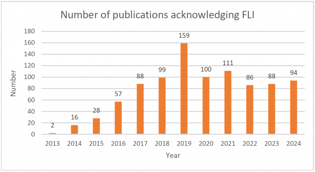
930 publications acknowledged FLI in the Web Of Science so far, 94 articles published in 2024, 88 published in 2023, 79 published in 2022, 110 published in 2021, 100 published in 2020, 159 published in 2019, 99 published in 2018, 89 published in 2017, 57 published in 2016, 39 published in 2015, 17 published in 2014 and 2 in 2013. Please find the complete list below.
2024
-
- F. Abbas, L. Blömer, H. Millet, J. Montnach, M. De Waard, et M. Canepari, « Analysis of the effect of the scorpion toxin AaH-II on action potential generation in the axon initial segment », SCIENTIFIC REPORTS, vol. 14, no 1, févr. 2024, doi: 10.1038/s41598-024-55315-y.
- M. Amouroux et al., « Multimodal characterization of optical properties of urinary stones ex vivo by machine-learning classification methods based on autofluorescence and integrating sphere measurements data: feasibility study & preliminary results », présenté à TISSUE OPTICS AND PHOTONICS III, V. Tuchin, W. Blondel, et Z. Zalevsky, Éd., 2024. doi: 10.1117/12.3016446.
- G. Andrade-Miranda, P. Vega, K. Taguelmimt, H. Dang, D. Visvikis, et J. Bert, « Exploring transformer reliability in clinically significant prostate cancer segmentation: A comprehensive in-depth investigation », COMPUTERIZED MEDICAL IMAGING AND GRAPHICS, vol. 118, déc. 2024, doi: 10.1016/j.compmedimag.2024.102459.
- A. Baldit et al., « Biomechanical Simulation of Haemostatic Sponges for Sinus Lift Application », présenté à COMPUTER METHODS IN BIOMECHANICS AND BIOMEDICAL ENGINEERING II, CMBBE 2023, S. Laporte, A. Benoit, et W. Skalli, Éd., 2024, p. 47‑57. doi: 10.1007/978-3-031-55315-8_6.
- G. Barone-Rochette, E. Lecesne, A. Simon, M. Garreau, et C. Fouard, « New Method CMR-Guided Endomyocardial Biopsy in Suspicion Context of Isolated Cardiac Sarcoidosis », CIRCULATION-CARDIOVASCULAR IMAGING, vol. 17, no 4, avr. 2024, doi: 10.1161/CIRCIMAGING.123.015807.
- L. Baynat et al., « Quantitative MRI Biomarkers Measure Changes in Targeted Brain Areas in Patients With Obesity », JOURNAL OF CLINICAL ENDOCRINOLOGY & METABOLISM, vol. 109, no 7, p. 1850‑1857, janv. 2024, doi: 10.1210/clinem/dgae014.
- Y. Becker et al., « Planum temporale asymmetry in newborn monkeys predicts the future development of gestural communication’s handedness », NATURE COMMUNICATIONS, vol. 15, no 1, juin 2024, doi: 10.1038/s41467-024-47277-6.
- L. Blömer et al., « Kinetics and functional consequences of BK channels activation by N-type Ca2+ channels in the dendrite of mouse neocortical layer-5 pyramidal neurons », FRONTIERS IN CELLULAR NEUROSCIENCE, vol. 18, févr. 2024, doi: 10.3389/fncel.2024.1353895.
- J. Boutin et al., « CRISPR editing to mimic porphyria combined with light: A new preclinical approach for prostate cancer », MOLECULAR THERAPY ONCOLOGY, vol. 32, no 1, mars 2024, doi: 10.1016/j.omton.2024.200772.
- C. Caredda et al., « A priori free spectral unmixing with periodic absorbance changes: application for auto-calibrated intraoperative functional brain mapping », BIOMEDICAL OPTICS EXPRESS, vol. 15, no 1, p. 387‑412, janv. 2024, doi: 10.1364/BOE.491292.
- C. Caredda et al., « Pixel-wise and real-time estimation of optical mean path length using deep learning: application for intraoperative functional brain mapping », présenté à CLINICAL BIOPHOTONICS III, S. Gioux, B. Pogue, et D. Elson, Éd., 2024. doi: 10.1117/12.3016632.
- C. Caredda, F. Lange, L. Giannoni, I. Ezhov, I. Tachtsidis, et B. Montcel, « A digital instrument simulator to optimize the development of hyperspectral systems for intraoperative brain mapping », présenté à TISSUE OPTICS AND PHOTONICS III, V. Tuchin, W. Blondel, et Z. Zalevsky, Éd., 2024. doi: 10.1117/12.3017488.
- Z. Chai et al., « Thieno3,2-b.thiophene-based bridged BODIPY dimers: synthesis, electrochemistry, and one- and two-photon photophysical properties », DALTON TRANSACTIONS, vol. 54, no 2, p. 674‑682, janv. 2025, doi: 10.1039/d4dt02655a.
- B. Chatel et al., « Cyclosporine A Delays the Terminal Disease Stage in the Tfam KO Mitochondrial Myopathy Mouse Model Without Improving Mitochondrial Energy Production », MUSCLE & NERVE, déc. 2024, doi: 10.1002/mus.28315.
- L. Chougar et al., « Contribution of MRI for the Early Diagnosis of Parkinsonism in Patients with Diagnostic Uncertainty », MOVEMENT DISORDERS, vol. 39, no 5, p. 825‑835, mai 2024, doi: 10.1002/mds.29760.
- C. Cornu et al., « CMUT for ultrafast passive cavitation detection during ultrasound-induced blood-brain barrier disruption: proof of concept study », PHYSICS IN MEDICINE AND BIOLOGY, vol. 69, no 20, oct. 2024, doi: 10.1088/1361-6560/ad8334.
- P. Daudé et al., « Trajectory correction enables free-running chemical shift encoded imaging for accurate cardiac proton-density fat fraction quantification at 3T », JOURNAL OF CARDIOVASCULAR MAGNETIC RESONANCE, vol. 26, no 2, WIN 2024, doi: 10.1016/j.jocmr.2024.101048.
- C. Delon-Martin et al., « Neural Correlates of Pain-Autonomic Coupling in Patients With Complex Regional Pain Syndrome Treated by Repetitive Transcranial Magnetic Stimulation of the Motor Cortex », NEUROMODULATION, vol. 27, no 1, p. 188‑199, janv. 2024, doi: 10.1016/j.neurom.2023.05.005.
- J. Deloulme et al., « Structural interhemispheric connectivity defects in mouse models of BBSOAS: Insights from high spatial resolution 3D white matter tractography », NEUROBIOLOGY OF DISEASE, vol. 193, avr. 2024, doi: 10.1016/j.nbd.2024.106455.
- T. Desbordes, J. King, et S. Dehaene, « Tracking the neural codes for words and phrases during semantic composition working-memory storage, and retrieval », CELL REPORTS, vol. 43, no 3, mars 2024, doi: 10.1016/j.celrep.2024.113847.
- L. Dietze et al., « White matter microstructure in obesity and bipolar disorders: an ENIGMA bipolar disorder working group study in 2186 individuals », MOLECULAR PSYCHIATRY, nov. 2024, doi: 10.1038/s41380-024-02784-2.
- J. Dillenseger et al., « Quantitative and qualitative evaluation of three MSCT for high resolution bone imaging », EUROPEAN JOURNAL OF RADIOLOGY, vol. 173, avr. 2024, doi: 10.1016/j.ejrad.2024.111394.
- P. Dodet et al., « ISOLATED REM SLEEP WITHOUT ATONIA IN EARLY-STAGE PARKINSON’S DISEASE IS NOT SYNONYMOUS OF REM SLEEP BEHAVIOR DISORDER », SLEEP MEDICINE, vol. 115, p. 303‑303, févr. 2024.
- P. Dodet et al., « SLEEP DISORDERS IN PARKINSON’S DISEASE, AN EARLY AND MULTIPLE PROBLEM », SLEEP MEDICINE, vol. 115, p. 202‑202, févr. 2024.
- P. Dodet et al., « Sleep disorders in Parkinson’s disease, an early and multiple problem », NPJ PARKINSONS DISEASE, vol. 10, no 1, févr. 2024, doi: 10.1038/s41531-024-00642-0.
- P. Dodet et al., « Isolated REM Sleep without Atonia Is Not Equivalent to REM Sleep Behavior Disorder in Early-Stage Parkinson’s Disease », MOVEMENT DISORDERS, vol. 39, no 7, p. 1190‑1202, juill. 2024, doi: 10.1002/mds.29813.
- T. Elsen et al., « A dataset of optical spectra and clinical features acquired on human healthy skin and on skin carcinomas », DATA IN BRIEF, vol. 53, avr. 2024, doi: 10.1016/j.dib.2024.110163.
- A. Fraissenon et al., « Sotorasib for Vascular Malformations Associated with KRAS G12C Mutation », NEW ENGLAND JOURNAL OF MEDICINE, vol. 391, no 4, p. 334‑342, juill. 2024, doi: 10.1056/NEJMoa2309160.
- G. Fu et al., « Projected pooling loss for red nucleus segmentation with soft topology constraints », JOURNAL OF MEDICAL IMAGING, vol. 11, no 4, juill. 2024, doi: 10.1117/1.JMI.11.4.044002.
- C. Fulbert et al., « Nanoscintillator Coating: A Key Parameter That Strongly Impacts Internalization, Biocompatibility, and Therapeutic Efficacy in Pancreatic Cancer Models », SMALL SCIENCE, vol. 4, no 5, mai 2024, doi: 10.1002/smsc.202400041.
- E. Gabory, A. Gautheron, M. Galtierb, M. Roger, et B. Montcel, « Estimation of 5-ALA-Induced PpIX Concentration in Fluorescence Spectroscopy for Improving Glioma Classification using Symbolic Monte Carlo », présenté à TISSUE OPTICS AND PHOTONICS III, V. Tuchin, W. Blondel, et Z. Zalevsky, Éd., 2024. doi: 10.1117/12.3017461.
- A. Garcia-Munoz, I. Varlet, G. Grau, T. Perles-Barbacaru, et A. Viola, « Contribution of Magnetic Resonance Imaging Studies to the Understanding of Cerebral Malaria Pathogenesis », PATHOGENS, vol. 13, no 12, déc. 2024, doi: 10.3390/pathogens13121042.
- A. Gautheron, J. Bernstock, T. Picart, J. Guyotat, P. Valdés, et B. Montcel, « 5-ALA induced PpIX fluorescence spectroscopy in neurosurgery: a review », FRONTIERS IN NEUROSCIENCE, vol. 18, janv. 2024, doi: 10.3389/fnins.2024.1310282.
- A. Gautheron, R. Clerc, V. Duveiller, L. Simonot, B. Montcel, et M. Hebert, « On the validity of two-flux and four-flux models for light scattering in translucent layers: angular distribution of internally reflected light at the interfaces », OPTICS EXPRESS, vol. 32, no 6, p. 9042‑9060, mars 2024, doi: 10.1364/OE.510888.
- A. Gautheron et al., « Robust estimation of 5-ALA-Induced PpIX Contributions in Multiple-Wavelength Excitation Fluorescence Spectroscopy to Improve Intraoperative Glioma Detection: Application on Clinical data », présenté à CLINICAL BIOPHOTONICS III, S. Gioux, B. Pogue, et D. Elson, Éd., 2024. doi: 10.1117/12.3022093.
- V. Gautier, A. Bousse, F. Sureau, C. Comtat, Maxim, et B. Sixou, « Bimodal PET/MRI generative reconstruction based on VAE architectures », PHYSICS IN MEDICINE AND BIOLOGY, vol. 69, no 24, déc. 2024, doi: 10.1088/1361-6560/ad9133.
- J. Gobé et al., « High-resolution brain tractography from X-ray phase-contrast images », EUROPEAN PHYSICAL JOURNAL PLUS, vol. 139, no 7, juill. 2024, doi: 10.1140/epjp/s13360-024-05357-y.
- E. Gonzalez et al., « Increased sighing during sleep as a marker of multiple system atrophy », NPJ PARKINSONS DISEASE, vol. 10, no 1, sept. 2024, doi: 10.1038/s41531-024-00765-4.
- S. Goutal et al., « Brain Glucose Metabolism as a Readout of the Central Nervous System Impact of Cigarette Smoke Exposure and Withdrawal and the Effects of NFL-101, as an Immune-Based Drug Candidate for Smoking Cessation Therapy », ACS CHEMICAL NEUROSCIENCE, vol. 15, no 13, p. 2520‑2531, juin 2024, doi: 10.1021/acschemneuro.4c00204.
- S. Gregoire, G. Laloy-Borgna, J. Aichele, F. Lemoult, et S. Catheline, « Flexural pulse wave velocity in blood vessels », JOURNAL OF THE ACOUSTICAL SOCIETY OF AMERICA, vol. 155, no 5, p. 2948‑2958, mai 2024, doi: 10.1121/10.0025855.
- S. Grimaldi et al., « Energetic dysfunction and iron overload in early Parkinson’s disease: Two distinct mechanisms? », PARKINSONISM & RELATED DISORDERS, vol. 124, juill. 2024, doi: 10.1016/j.parkreldis.2024.106996.
- T. Hähnel et al., « Progression subtypes in Parkinson’s disease identified by a data-driven multi cohort analysis », NPJ PARKINSONS DISEASE, vol. 10, no 1, mai 2024, doi: 10.1038/s41531-024-00712-3.
- S. Harquel et al., « Modulation of Visually Induced Self-motion Illusions by α Transcranial Electric Stimulation over the Superior Parietal Cortex », JOURNAL OF COGNITIVE NEUROSCIENCE, vol. 36, no 1, p. 143‑154, janv. 2024, doi: 10.1162/jocn_a_02074.
- M. Hautiere et al., « Preoperative PET imaging and fluorescence-guided surgery of human glioblastoma using dual-labeled antibody targeting ETA receptors in a preclinical mouse model: A theranostic approach », THERANOSTICS, vol. 14, no 16, p. 6268‑6280, 2024, doi: 10.7150/thno.98163.
- Z. He, P. Soullié, P. Lefebvre, K. Ambarki, J. Felblinger, et F. Odille, « Changes of in vivo electrical conductivity in the brain and torso related to age, fat fraction and sex using MRI », SCIENTIFIC REPORTS, vol. 14, no 1, juill. 2024, doi: 10.1038/s41598-024-67014-9.
- R. Hervochon et al., « Cerebral Plasticity after Lengthening Temporalis Myoplasty in Facial Palsy: A Magnetoencephalography Study », FACIAL PLASTIC SURGERY & AESTHETIC MEDICINE, mai 2024, doi: 10.1089/fpsam.2023.0235.
- B. Hollunder et al., « Mapping dysfunctional circuits in the frontal cortex using deep brain stimulation », NATURE NEUROSCIENCE, vol. 27, no 3, p. 573‑586, mars 2024, doi: 10.1038/s41593-024-01570-1.
- B. Hosten et al., « Brain delivery enabled by transient blood-brain barrier disruption induced by regadenoson: a PET imaging study », EXPERT OPINION ON DRUG DELIVERY, vol. 21, no 5, p. 797‑807, mai 2024, doi: 10.1080/17425247.2024.2369765.
- K. Isaieva et al., « Extraction of 3D trajectories of mandibular condyles from 2D real-time MRI », MAGNETIC RESONANCE MATERIALS IN PHYSICS BIOLOGY AND MEDICINE, déc. 2024, doi: 10.1007/s10334-024-01214-2.
- O. Ishak et al., « Magnetic resonance cavitation imaging for the monitoring of ultrasound therapies », PHYSICS IN MEDICINE AND BIOLOGY, vol. 69, no 21, nov. 2024, doi: 10.1088/1361-6560/ad84b4.
- M. Joliot, S. Cremona, C. Tzourio, et O. Etard, « Modulate the impact of the drowsiness on the resting state functional connectivity », SCIENTIFIC REPORTS, vol. 14, no 1, avr. 2024, doi: 10.1038/s41598-024-59476-8.
- K. Jouad et al., « Multimodal PET/PAI/FLI imaging probe based on meso-O-alkyl heptamethine cyanine: Synthesis, 18F.F-radiolabelling and photophysical characterizations », DYES AND PIGMENTS, vol. 223, avr. 2024, doi: 10.1016/j.dyepig.2024.111995.
- S. Krystal et al., « Functional connectivity of the amygdala subnuclei in various mood states of bipolar disorder », MOLECULAR PSYCHIATRY, vol. 29, no 11, p. 3344‑3355, nov. 2024, doi: 10.1038/s41380-024-02580-y.
- V. Kupriyanov et al., « Bimodal spectroscopy for skin carcinomas and actinic keratoses diagnostic assistance », présenté à CLINICAL BIOPHOTONICS III, S. Gioux, B. Pogue, et D. Elson, Éd., 2024. doi: 10.1117/12.3016431.
- V. Kupriyanov et al., « Estimation of skin age and phototype using bimodal spectroscopy and machine learning », présenté à TISSUE OPTICS AND PHOTONICS III, V. Tuchin, W. Blondel, et Z. Zalevsky, Éd., 2024. doi: 10.1117/12.3016417.
- A. Lacroix et al., « Reduced spatial frequency differentiation and sex-related specificities in fearful face detection in autism: Insights from EEG and the predictive brain model », AUTISM RESEARCH, vol. 17, no 9, p. 1778‑1795, sept. 2024, doi: 10.1002/aur.3209.
- A. Lacroix et al., « Sex modulation of faces prediction error in the autistic brain », COMMUNICATIONS BIOLOGY, vol. 7, no 1, janv. 2024, doi: 10.1038/s42003-024-05807-4.
- S. Lee et al., « A Deep Learning Approach for Placing Magnetic Resonance Spectroscopy Voxels in Brain Tumors », présenté à MEDICAL IMAGE COMPUTING AND COMPUTER ASSISTED INTERVENTION – MICCAI 2024, PT III, A. Feragen, S. Giannarou, B. Glocker, K. Lekadir, J. Schnabel, M. Linguraru, et Q. Dou, Éd., 2024, p. 543‑552. doi: 10.1007/978-3-031-72384-1_51.
- S. Leterrier et al., « Imaging quantitative changes in blood-brain barrier permeability using 18F.2-fluoro-2-deoxy-sorbitol (18F.FDS) PET in relation to glial cell recruitment in a mouse model of endotoxemia », JOURNAL OF CEREBRAL BLOOD FLOW AND METABOLISM, vol. 44, no 7, p. 1117‑1127, juill. 2024, doi: 10.1177/0271678X241236755.
- C. Linger, G. Maccini, G. Clavier, R. Méallet, N. Tsapis, et J. Gateau, « Quantitative photoacoustic spectral transformations in theranostic solid lipid nanoparticles labelled with increasing concentrations of a photoacoustic NIR BODIPY », NANOSCALE, vol. 17, no 1, p. 440‑458, déc. 2024, doi: 10.1039/d4nr02880e.
- H. Lokossou, G. Rabuffo, M. Bernard, C. Bernard, et T. Perles-Barbacaru, « Impact of the day/night cycle on functional connectome in ageing male and female mice », NEUROIMAGE, vol. 290, avr. 2024, doi: 10.1016/j.neuroimage.2024.120576.
- V. López-Madrona et al., « Identification of Early Hippocampal Dynamics during Recognition Memory with Independent Component Analysis », ENEURO, vol. 11, no 4, avr. 2024, doi: 10.1523/ENEURO.0183-23.2023.
- Z. Lyu et al., « Self- assembling dendrimer nanosystems for specific fluorine magnetic resonance imaging and effective theranostic treatment of tumors », PROCEEDINGS OF THE NATIONAL ACADEMY OF SCIENCES OF THE UNITED STATES OF AMERICA, vol. 121, no 25, juin 2024, doi: 10.1073/pnas.2322403121.
- S. Marhabaie, A. Labbé, B. Quesson, et M. Poirier-Quinot, « The Minimum Admissible Detuning Efficiency of MRI Receive-Only Surface Coils », JOURNAL OF MAGNETIC RESONANCE IMAGING, vol. 60, no 2, p. 777‑788, août 2024, doi: 10.1002/jmri.29209.
- A. Marmin, N. Dufour, S. Facca, S. Catheline, S. Chatelin, et A. Nahas, « Full-field noise-correlation elastography for in-plane mechanical anisotropy imaging », BIOMEDICAL OPTICS EXPRESS, vol. 15, no 4, p. 2622‑2635, avr. 2024, doi: 10.1364/BOE.516166.
- S. Meoni, M. Dojat, M. Hutchinson, P. Pelissier, C. Chiquet, et E. Moro, « Visual dysfunction of superior colliculus and lateral geniculate nucleus in idiopathic blepharospasm », JOURNAL OF THE NEUROLOGICAL SCIENCES, vol. 466, nov. 2024, doi: 10.1016/j.jns.2024.123272.
- C. Michel et al., « Endurance training and hydroxyurea have synergistic effects on muscle function and energetics in sickle cell disease mice », BLOOD CELLS MOLECULES AND DISEASES, vol. 107, juill. 2024, doi: 10.1016/j.bcmd.2024.102853.
- K. Mishima et al., « Quantification of hemi-hepatic ischemia using real-time multispectral oxygenation imaging with single snapshot imaging of optical properties (SSOP) », SURGICAL ENDOSCOPY AND OTHER INTERVENTIONAL TECHNIQUES, déc. 2024, doi: 10.1007/s00464-024-11435-0.
- B. Montcel, C. Caredda, et P. Valdés, « Editorial: Advancements in intraoperative optical technologies for neurosurgery guidance », FRONTIERS IN NEUROSCIENCE, vol. 18, déc. 2024, doi: 10.3389/fnins.2024.1527174.
- J. Mutin et al., « Validation of HPLC and TLC analytical methods to determine radiochemical purity of 99m Tc-cAbVCAM1-5, a new experimental radiotracer », JOURNAL OF PHARMACEUTICAL AND BIOMEDICAL ANALYSIS, vol. 246, août 2024, doi: 10.1016/j.jpba.2024.116224.
- H. Ngo et al., « In plane quantification of in vivo muscle elastic anisotropy factor by steered ultrasound pushing beams », PHYSICS IN MEDICINE AND BIOLOGY, vol. 69, no 4, févr. 2024, doi: 10.1088/1361-6560/ad21a0.
- P. Nobre, G. Gaborit, A. Troia, U. Zanovello, L. Duvillaret, et O. Beuf, « Electric field measurements in preclinical MRI at 11.7 T and 7 T for experimental SAR comparison », JOURNAL OF MAGNETISM AND MAGNETIC MATERIALS, vol. 593, mars 2024, doi: 10.1016/j.jmmm.2024.171818.
- A. Omouri, S. Rapacchi, J. Duclos, R. Niddam, M. Bellemare, et N. Pirró, « 3D Observation of Pelvic Organs with Dynamic MRI Segmentation: A Bridge Toward Patient-Specific Models », INTERNATIONAL UROGYNECOLOGY JOURNAL, vol. 35, no 7, p. 1389‑1397, juill. 2024, doi: 10.1007/s00192-024-05817-0.
- T. Picart et al., « Fluorescence-Guided Surgical Techniques in Adult Diffuse Low-Grade Gliomas: State-of-the-Art and Emerging Techniques: A Systematic Review », CANCERS, vol. 16, no 15, août 2024, doi: 10.3390/cancers16152698.
- M. Potez et al., « Microbeam Radiation Therapy Opens a Several Days’ ’ Vessel Permeability Window for Small Molecules in Brain Tumor Vessels », INTERNATIONAL JOURNAL OF RADIATION ONCOLOGY BIOLOGY PHYSICS, vol. 119, no 5, p. 1506‑1516, août 2024, doi: 10.1016/j.ijrobp.2024.02.007.
- H. Quelquejay et al., « L-Wnk1 Deletion in Smooth Muscle Cells Causes Aortitis and Inflammatory Shift », CIRCULATION RESEARCH, vol. 135, no 4, p. 488‑502, août 2024, doi: 10.1161/CIRCRESAHA.124.324366.
- A. Razzouki et al., « Early-Stage Parkinson’s Disease Detection Based on Optical Flow and Video Vision Transformer », présenté à 2024 16TH INTERNATIONAL CONFERENCE ON HUMAN SYSTEM INTERACTION, HSI 2024, 2024. doi: 10.1109/HSI61632.2024.10613585.
- S. Rigollet et al., « FUS-mediated BBB opening leads to transient perfusion decrease and inflammation without acute or chronic brain lesion », THERANOSTICS, vol. 14, no 10, p. 4147‑4160, 2024, doi: 10.7150/thno.96721.
- P. Robert et al., « Auditory hemispheric asymmetry for actions and objects », CEREBRAL CORTEX, vol. 34, no 7, juill. 2024, doi: 10.1093/cercor/bhae292.
- S. Rodrigo et al., « Brain 18 F-FDG PET reveals cortico-subcortical hypermetabolic dysfunction in juvenile neuropsychiatric systemic lupus erythematosus », EJNMMI RESEARCH, vol. 14, no 1, avr. 2024, doi: 10.1186/s13550-024-01088-4.
- L. Roncali et al., « Brain intratumoural astatine-211 radiotherapy targeting syndecan-1 leads to durable glioblastoma remission and immune memory in female mice », EBIOMEDICINE, vol. 105, juill. 2024, doi: 10.1016/j.ebiom.2024.105202.
- T. Roussel et al., « Impact of inner hydrophobicity of dendrimer nanomicelles on biodistribution: a PET imaging study », JOURNAL OF MATERIALS CHEMISTRY B, déc. 2024, doi: 10.1039/d4tb01266f.
- A. Sadikine et al., « Improving abdominal image segmentation with overcomplete shape priors », COMPUTERIZED MEDICAL IMAGING AND GRAPHICS, vol. 113, avr. 2024, doi: 10.1016/j.compmedimag.2024.102356.
- P. Sango-Solanas et al., « Ultrashort echo time magnetic resonance elastography for quantification of the mechanical properties of short T2 tissues via optimal control-based radiofrequency pulses », NMR IN BIOMEDICINE, vol. 37, no 11, nov. 2024, doi: 10.1002/nbm.5210.
- A. Serrurier et C. Neuschaefer-Rube, « Formant-based articulatory strategies: Characterisation and inter-speaker variability analysis », JOURNAL OF PHONETICS, vol. 107, nov. 2024, doi: 10.1016/j.wocn.2024.101374.
- A. Simon et al., « Expanding the Palette of SWIR Emitting Nanoparticles Based on Au Nanoclusters for Single-Particle Tracking Microscopy », ADVANCED SCIENCE, vol. 11, no 24, juin 2024, doi: 10.1002/advs.202309267.
- A. Soyer et al., « 18F.2-fluoro-2-deoxy-sorbitol (18F.FDS) PET imaging repurposed for quantitative estimation of blood-brain barrier permeability in a rat model of Alzheimer’s disease », ANNALES PHARMACEUTIQUES FRANCAISES, vol. 82, no 5, p. 822‑829, sept. 2024, doi: 10.1016/j.pharma.2024.04.004.
- P. Stenroos et al., « EEG-fMRI in awake rat and whole-brain simulations show decreased brain responsiveness to sensory stimulations during absence seizures », ELIFE, vol. 12, juill. 2024, doi: 10.7554/eLife.90318.
- A. Szczodra et al., « Boron substitution in silicate bioactive glass scaffolds to enhance bone differentiation and regeneration », ACTA BIOMATERIALIA, vol. 186, p. 489‑506, sept. 2024, doi: 10.1016/j.actbio.2024.07.053.
- B. Testud, X. Carle, C. Costes, J. Hak, et M. Guye, « Added Value of Ultrahigh-Resolution 7T MRI in Dural Arteriovenous Fistulas », STROKE, vol. 55, no 3, p. E46‑E47, mars 2024, doi: 10.1161/STROKEAHA.123.045930.
- S. Todorovic et al., « Cortico-Cerebellar Monitoring of Speech Sequence Production », NEUROBIOLOGY OF LANGUAGE, vol. 5, no 3, p. 701‑721, août 2024, doi: 10.1162/nol_a_00113.
- A. Uzel, M. Sdika, S. Chopinet, O. Lopez, et B. Montcel, « Near Infrared Diffuse Reflectance Spectroscopy for fat quantification in Fatty Liver Disease », présenté à ADVANCED BIOMEDICAL AND CLINICAL DIAGNOSTIC AND SURGICAL GUIDANCE SYSTEMS XXII, C. Boudoux et J. Tunnell, Éd., 2024. doi: 10.1117/12.3001895.
- C. Vazia et al., « Spectral CT Two-step and One-step Material Decomposition using Diffusion Posterior Sampling », présenté à 32ND EUROPEAN SIGNAL PROCESSING CONFERENCE, EUSIPCO 2024, 2024, p. 1506‑1510.
94. L. Zerbib et al., « Targeted therapy for capillary-venous malformations », SIGNAL TRANSDUCTION AND TARGETED THERAPY, vol. 9, no 1, juin 2024, doi: 10.1038/s41392-02
2023
- Achard, S.; Coeurjolly, J.-F.; de Micheaux, P. L.; Lbath, H.; Richiardi, J. Inter-Regional Correlation Estimators for Functional Magnetic Resonance Imaging. NEUROIMAGE 2023, 282. https://doi.org/10.1016/j.neuroimage.2023.120388.
- Andrade-Miranda, G.; Jaouen, V.; Tankyevych, O.; Le Rest, C. C.; Visvikis, D.; Conze, P.-H. Multi-Modal Medical Transformers: A Meta-Analysis for Medical Image Segmentation in Oncology. COMPUTERIZED MEDICAL IMAGING AND GRAPHICS 2023, 110. https://doi.org/10.1016/j.compmedimag.2023.102308.
- Badier, J.-M.; Schwartz, D.; Benar, C.-G.; Kanzari, K.; Daligault, S.; Romain, R.; Mitryukovskiy, S.; Fourcault, W.; Josselin, V.; Le Prado, M.; Jung, J.; Palacios-Laloy, A.; Roman, C.; Bartolomei, F.; Laby, E.; Bonini, F. Helium Optically Pumped Magnetometers Can Detect Epileptic Abnormalities as Well as SQUIDs as Shown by Intracerebral Recordings. ENEURO 2023, 10 (12). https://doi.org/10.1523/ENEURO.0222-23.2023.
- Bailly, T.; Bodin, S.; Goncalves, V.; Denat, F.; Morgat, C.; Prignon, A.; Valverde, I. E. Modular One-Pot Strategy for the Synthesis of Heterobivalent Tracers. ACS MEDICINAL CHEMISTRY LETTERS 2023, 14 (5), 636–644. https://doi.org/10.1021/acsmedchemlett.3c00057.
- Bayard, C.; Segna, E.; Taverne, M.; Fraissenon, A.; Hennocq, Q.; Periou, B.; Zerbib, L.; Ladraa, S.; Chapelle, C.; Hoguin, C.; Kaltenbach, S.; Villarese, P.; Asnafi, V.; Broissand, C.; Nemazanyy, I.; Autret, G.; Goudin, N.; Legendre, C.; Authier, F.-J.; Viel, T.; Tavitian, B.; Gitiaux, C.; Fraitag, S.; Duong, J.-P.; Delcros, C.; Sergent, B.; Picard, A.; Dussiot, M.; Guibaud, L.; Khonsari, R.; Canaud, G. Hemifacial Myohyperplasia Is Due to Somatic Muscular PIK3CA Gain-of-Function Mutations and Responds to Pharmacological Inhibition. JOURNAL OF EXPERIMENTAL MEDICINE 2023, 220 (11). https://doi.org/10.1084/jem.20230926.
- Bertho, A.; Iturri, L.; Brisebard, E.; Juchaux, M.; Gilbert, C.; Ortiz, R.; Sebrie, C.; Jourdain, L.; Lamirault, C.; Ramasamy, G.; Pouzoulet, F.; Prezado, Y. Evaluation of the Role of the Immune System Response After Minibeam Radiation Therapy. INTERNATIONAL JOURNAL OF RADIATION ONCOLOGY BIOLOGY PHYSICS 2023, 115 (2), 426–439.
- Bery, A.; Etienne, O.; Mouton, L.; Mokrani, S.; Granotier-Beckers, C.; Gauthier, L. R.; Feat-Vetel, J.; Kortulewski, T.; Peres, E. A.; Desmaze, C.; Lestaveal, P.; Barroca, V.; Laugeray, A.; Boumezbeur, F.; Abramovski, V.; Mortaud, S.; Menuet, A.; Le Bihan, D.; de Villartay, J.-P.; Boussin, F. D. XLF/Cernunnos Loss Impairs Mouse Brain Development by Altering Symmetric Proliferative Divisions of Neural Progenitors. CELL REPORTS 2023, 42 (4). https://doi.org/10.1016/j.celrep.2023.112342.
- Bodin, S.; Previti, S.; Jestin, E.; Vimont, D.; Ait-Arsa, I.; Lamare, F.; Remond, E.; Hindie, E.; Cavelier, F.; Morgat, C. Design, Synthesis, and Biological Evaluation of the First Radio- Metalated Neurotensin Analogue Targeting Neurotensin Receptor 2. ACS OMEGA 2023. https://doi.org/10.1021/acsomega.2c07814.
- Bodin, S.; Previti, S.; Jestin, E.; Vimont, D.; Ait-Arsa, I.; Remond, E.; Hindie, E.; Cavelier, F.; Morgat, C.; Lamare, F. Design, Synthesis, and Biological Evaluation of the First Radio-Metalated Neurotensin Analogue Targeting Neurotensin Receptor 2. ACS OMEGA 2023. https://doi.org/10.1021/acsomega.2c07814.
- Bourdin, V.; Charlier, P.; Crevat, S.; Slimani, L.; Chaussain, C.; Kielbasa, M.; Pible, O.; Armengaud, J. Deep Paleoproteotyping and Microtomography Revealed No Heart Defect nor Traces of Embalming in the Cardiac Relics of Blessed Pauline Jaricot. INTERNATIONAL JOURNAL OF MOLECULAR SCIENCES 2023, 24 (3). https://doi.org/10.3390/ijms24033011.
- Branzoli, F.; Liserre, R.; Deelchand, D. K.; Poliani, P. L.; Bielle, F.; Nichelli, L.; Sanson, M.; Lehericy, S.; Marjanska, M. Neurochemical Differences between 1p/19q Codeleted and Noncodeleted IDH-Mutant Gliomas by in Vivo MR Spectroscopy. RADIOLOGY 2023, 308 (3). https://doi.org/10.1148/radiol.223255.
- Branzoli, F.; Salgues, B.; Marjanska, M.; Laloi-Michelin, M.; Herman, P.; Le Collen, L.; Delemer, B.; Riancho, J.; Kuhn, E.; Jublanc, C.; Burnichon, N.; Amar, L.; Favier, J.; Gimenez-Roqueplo, A.-P.; Buffet, A.; Lussey-Lepoutre, C. SDHx Mutation and Pituitary Adenoma: Can in Vivo <SUP>1</SUP>H-MR Spectroscopy Unravel the Link? ENDOCRINE-RELATED CANCER 2023, 30 (2). https://doi.org/10.1530/ERC-22-0198.
- Caredda, C.; Cohen, J. E.; Mahieu-Williame, L.; Sablong, R.; Sdika, M.; Guyotat, J.; Montcel, B. Separable Spectral Unmixing Based on the Learning of Periodic Absorbance Changes: Application to Functional Brain Mapping Using RGB Imaging. In TRANSLATIONAL BIOPHOTONICS: DIAGNOSTICS AND THERAPEUTICS III; Huang, Z., Lilge, L., Eds.; Proceedings of SPIE; SPIE; Optica, 2023; Vol. 12627. https://doi.org/10.1117/12.2670506.
- Casali, V.; Guithon, I. C.; Sanden, B. van der; Stephan, O.; Gredy, L.; Vilgrain, I.; Martin, D. K. Is a Real-Time Quantifiable Liquid Biopsy Achievable Using a Microfluidic Lab-on-Chip ? EUROBIOTECH JOURNAL 2023, 7 (4), 189–195. https://doi.org/10.2478/ebtj-2023-0014.
- Chevaleyre, C.; Novell, A.; Tournier, N.; Dauba, A.; Dubois, S.; Kereselidze, D.; Selingue, E.; Jego, B.; Maillere, B.; Larrat, B.; Nozach, H.; Truillet, C. Efficient PD-L1 Imaging of Murine Glioblastoma with FUS-Aided ImmunoPET by Leveraging FcRn-Antibody Interaction. THERANOSTICS 2023, 13 (15), 5584–5596. https://doi.org/10.7150/thno.87168.
- Chougar, L.; Lejeune, F.-X.; Faouzi, J.; Morino, B.; Faucher, A.; Hoyek, N.; Grabli, D.; Cormier, F.; Vidailhet, M.; Corvol, J.-C.; Colliot, O.; Degos, B.; Lehericy, S. Comparison of Mean Diffusivity, R2*relaxation Rate and Morphometric Biomarkers for the Clinical Differentiation of Parkinsonism. PARKINSONISM & RELATED DISORDERS 2023, 108. https://doi.org/10.1016/j.parkreldis.2023.105287.
- Commowick, O.; Combes, B.; Cervenansky, F.; Dojat, M. Editorial: Automatic Methods for Multiple Sclerosis New Lesions Detection and Segmentation. FRONTIERS IN NEUROSCIENCE 2023, 17. https://doi.org/10.3389/fnins.2023.1176625.
- Conze, P.-H.; Andrade-Miranda, G.; Singh, V. K.; Jaouen, V.; Visvikis, D. Current and Emerging Trends in Medical Image Segmentation With Deep Learning. IEEE TRANSACTIONS ON RADIATION AND PLASMA MEDICAL SCIENCES 2023, 7 (6), 545–569. https://doi.org/10.1109/TRPMS.2023.3265863.
- Courtin, C.; Lacoin, G.; Remenieras, J.-P.; Rousselot, C. D.; Dujardin, P.-A.; Zemmoura, I.; Cottier, J.-P. Tumoral and Peritumoral Vascularization of Brain Tumours: A Study Comparing an Intraoperative Ultrasensitive Doppler and a Preoperative First-Pass Perfusion MRI. NEUROCHIRURGIE 2023, 69 (6). https://doi.org/10.1016/j.neuchi.2023.101493.
- Daude, P.; Roussel, T.; Troalen, T.; Viout, P.; Hernando, D.; Guye, M.; Kober, F.; Gouny, S. C.; Bernard, M.; Rapacchi, S. Comparative Review of Algorithms and Methods for Chemical-Shift-Encoded Quantitative Fat-Water Imaging. MAGNETIC RESONANCE IN MEDICINE 2023. https://doi.org/10.1002/mrm.29860.
- Davin, A.; Chabardes, S.; Devergnas, A.; Benstaali, C.; Gutekunst, C.-A. N.; David, O.; Torres-Martinez, N.; Piallat, B. Excessive Daytime Sleepiness in a Model of Parkinson’s Disease Improved by Low-Frequency Stimulation of the Pedunculopontine Nucleus. NPJ PARKINSONS DISEASE 2023, 9 (1). https://doi.org/10.1038/s41531-023-00455-7.
- de Souza, D. A. R.; Mathieu, H.; Deloulme, J.-C.; Barbier, E. L. Evaluation of Kernel Low-Rank Compressed Sensing in Preclinical Diffusion Magnetic Resonance Imaging. FRONTIERS IN NEUROSCIENCE 2023, 17. https://doi.org/10.3389/fnins.2023.1172830.
- Desbordes, T.; Lakretz, Y.; Chanoine, V.; Oquab, M.; Badier, J.-M.; Trebuchon, A.; Carron, R.; Benar, C.-G.; Dehaene, S.; King, J.-R. Dimensionality and Ramping: Signatures of Sentence Integration in the Dynamics of Brains and Deep Language Models. JOURNAL OF NEUROSCIENCE 2023, 43 (29), 5350–5364. https://doi.org/10.1523/JNEUROSCI.1163-22.2023.
- Destruel, A.; Mauconduit, F.; Massire, A.; Abdeddaim, R.; Guye, M.; Gras, V.; Callot, V. Optimized Interferometric Encoding of Presaturated TurboFLASH B1 Mapping for Parallel Transmission MRI at 7 T: Preliminary Application for Quantitative T1 Mapping in the Spinal Cord. MAGNETIC RESONANCE IN MEDICINE 2023. https://doi.org/10.1002/mrm.29708.
- Ding, L.; Lyu, Z.; Perles-Barbacaru, T.-A.; Huang, A. Y.-T.; Lian, B.; Jiang, Y.; Roussel, T.; Galanakou, C.; Giorgio, S.; Kao, C.-L.; Liu, X.; Iovanna, J.; Bernard, M.; Viola, A.; Peng, L. Modular Self-Assembling Dendrimer Nanosystems for Magnetic Resonance and Multimodality Imaging of Tumors. ADVANCED MATERIALS 2023. https://doi.org/10.1002/adma.202308262.
- dos Santos, E. J. L.; Nakajima, K.; Po, J.; Hanai, A.; Zhukouskaya, V.; Duplan, M. B.; Linglart, A.; Shimada, T.; Chaussain, C.; Bardet, C. Dental Impact of Anti-Fibroblast Growth Factor 23 Therapy in X-Linked Hypophosphatemia. INTERNATIONAL JOURNAL OF ORAL SCIENCE 2023, 15 (1). https://doi.org/10.1038/s41368-023-00259-8.
- Filipis, L.; Blomer, L. A.; Montnach, J.; Loussouarn, G.; De Waard, M.; Canepari, M. Nav1.2 and BK Channel Interaction Shapes the Action Potential in the Axon Initial Segment. JOURNAL OF PHYSIOLOGY-LONDON 2023, 601 (10), 1957–1979. https://doi.org/10.1113/JP283801.
- Echavidre W.; Durivault J.; Gotorbe C.; Blanchard T.; Pagnuzzi M.; Vial V.;, Raes F.; Broisat A.; Villeneuve R.; Amblard R.; Garnier N.; Ortholan C.; Faraggi M.; Serrano B.; Picco V.; and Montemagno C. Integrin-αvβ3 is a therapeutically targetable fundamental factor in medulloblastoma tumorigenicity and radioresistance. CANCER RESEARCH COMMUNICATIONS 2023. https://doi.org/10.1158/2767-9764.CRC-23-0298
- Fu, G.; El Jurdi, R.; Chougar, L.; Dormont, D.; Valabregue, R.; Lehericy, S.; Colliot, O.; Grp, I. S. Introducing Soft Topology Constraints in Deep Learning-Based Segmentation Using Projected Pooling Loss. In MEDICAL IMAGING 2023; Colliot, O., Isgum, I., Eds.; Progress in Biomedical Optics and Imaging; SPIE; Philips Healthcare, 2023; Vol. 12464. https://doi.org/10.1117/12.2651576.
- Garcelon, C.; Abascal, J.; Olivier, C.; Uk, S.; Si-Mohamed, S.; Ea, H.-K.; Douek, P.; Peyrin, F.; Chappard, C. Quantification of Cartilage and Subchondral Bone Cysts on Knee Specimens Based on a Spectral Photon-Counting Computed Tomography. SCIENTIFIC REPORTS 2023, 13 (1). https://doi.org/10.1038/s41598-023-38238-y.
- Garot, C.; Schoffit, S.; Monfoulet, C.; Machillot, P.; Deroy, C.; Roques, S.; Vial, J.; Vollaire, J.; Renard, M.; Ghanem, H.; El-Hafci, H.; Decambron, A.; Josserand, V.; Bordenave, L.; Bettega, G.; Durand, M.; Manassero, M.; Viateau, V.; Logeart-Avramoglou, D.; Picart, C. 3D-Printed Osteoinductive Polymeric Scaffolds with Optimized Architecture to Repair a Sheep Metatarsal Critical-Size Bone Defect. ADVANCED HEALTHCARE MATERIALS 2023. https://doi.org/10.1002/adhm.202301692.
- Goutal, S.; Novell, A.; Leterrier, S.; Breuil, L.; Selingue, E.; Gerstenmayer, M.; Marie, S.; Saubamea, B.; Caille, F.; Langer, O.; Truillet, C.; Larrat, B.; Tournier, N. Imaging the Impact of Blood-Brain Barrier Disruption Induced by Focused Ultrasound on P-Glycoprotein Function. JOURNAL OF CONTROLLED RELEASE 2023, 361, 483–492. https://doi.org/10.1016/j.jconrel.2023.08.012.
- Grandjean, J.; Desrosiers-Gregoire, G.; Anckaerts, C.; Angeles-Valdez, D.; Ayad, F.; Barriere, D. A.; Blockx, I.; Bortel, A.; Broadwater, M.; Cardoso, B. M.; Celestine, M.; Chavez-Negrete, J. E.; Choi, S.; Christiaen, E.; Clavijo, P.; Colon-Perez, L.; Cramer, S.; Daniele, T.; Dempsey, E.; Diao, Y.; Doelemeyer, A.; Dopfel, D.; Dvorakova, L.; Falfan-Melgoza, C.; Fernandes, F. F.; Fowler, C. F.; Fuentes-Ibanez, A.; Garin, C.; Gelderman, E.; Golden, C. E. M.; Guo, C. C. G.; Henckens, M. J. A. G.; Hennessy, L. A.; Herman, P.; Hofwijks, N.; Horien, C.; Ionescu, T. M.; Jones, J.; Kaesser, J.; Kim, E.; Lambers, H.; Lazari, A.; Lee, S.-H.; Lillywhite, A.; Liu, Y.; Liu, Y. Y.; Lopez-Castro, A.; Lopez-Gil, X.; Ma, Z.; MacNicol, E.; Madularu, D.; Mandino, F.; Marciano, S.; McAuslan, M. J.; McCunn, P.; McIntosh, A.; Meng, X.; Meyer-Baese, L.; Missault, S.; Moro, F.; Naessens, D. M. P.; Nava-Gomez, L. J.; Nonaka, H.; Ortiz, J. J.; Paasonen, J.; Peeters, L. M.; Pereira, M.; Perez, P. D.; Pompilus, M.; Prior, M.; Rakhmatullin, R.; Reimann, H. M.; Reinwald, J.; Del Rio, R. T.; Rivera-Olvera, A.; Ruiz-Perez, D.; Russo, G.; Rutten, T. J.; Ryoke, R.; Sack, M.; Salvan, P.; Sanganahalli, B. G.; Schroeter, A.; Seewoo, B. J.; Selingue, E.; Seuwen, A.; Shi, B.; Sirmpilatze, N.; Smith, J. A.; Smith, C.; Sobczak, F.; Stenroos, P. J.; Straathof, M.; Strobelt, S.; Sumiyoshi, A.; Takahashi, K.; Torres-Garcia, M. E.; Tudela, R.; van den Berg, M.; van der Marel, K.; van Hout, A. T. B.; Vertullo, R.; Vidal, B.; Vrooman, R. M.; Wang, V. X.; Wank, I.; Watson, D. J. G.; Yin, T.; Zhang, Y.; Zurbruegg, S.; Achard, S.; Alcauter, S.; Auer, D. P.; Barbier, E. L.; Baudewig, J.; Beckmann, C. F.; Beckmann, N.; Becq, G. J. P. C.; Blezer, E. L. A.; Bolbos, R.; Boretius, S.; Bouvard, S.; Budinger, E.; Buxbaum, J. D.; Cash, D.; Chapman, V.; Chuang, K.-H.; Ciobanu, L.; Coolen, B. F.; Dalley, J. W.; Dhenain, M.; Dijkhuizen, R. M.; Esteban, O.; Faber, C.; Febo, M.; Feindel, K. W.; Forloni, G.; Fouquet, J.; Garza-Villarreal, E. A.; Gass, N.; Glennon, J. C.; Gozzi, A.; Grohn, O.; Harkin, A.; Heerschap, A.; Helluy, X.; Herfert, K.; Heuser, A.; Homberg, J. R.; Houwing, D. J.; Hyder, F.; Ielacqua, G. D.; Jelescu, I. O.; Johansen-Berg, H.; Kaneko, G.; Kawashima, R.; Keilholz, S. D.; Keliris, G. A.; Kelly, C.; Kerskens, C.; Khokhar, J. Y.; Kind, P. C.; Langlois, J.-B.; Lerch, J. P.; Lopez-Hidalgo, M. A.; Manahan-Vaughan, D.; Marchand, F.; Mars, R. B.; Marsella, G.; Micotti, E.; Munoz-Moreno, E.; Near, J.; Niendorf, T.; Otte, W. M.; Pais-Roldan, P.; Pan, W.-J.; Prado-Alcala, R. A.; Quirarte, G. L.; Rodger, J.; Rosenow, T.; Sampaio-Baptista, C.; Sartorius, A.; Sawiak, S. J.; Scheenen, T. W. J.; Shemesh, N.; Shih, Y.-Y. I.; Shmuel, A.; Soria, G.; Stoop, R.; Thompson, G. J.; Till, S. M.; Todd, N.; van der Linden, A.; van der Toorn, A.; van Tilborg, G. A. F.; Vanhove, C.; Veltien, A.; Verhoye, M.; Wachsmuth, L.; Weber-Fahr, W.; Wenk, P.; Yu, X.; Zerbi, V.; Zhang, N.; Zhang, B. B.; Zimmer, L.; Devenyi, G. A.; Chakravarty, M. M.; Hess, A. A Consensus Protocol for Functional Connectivity Analysis in the Rat Brain. NATURE NEUROSCIENCE 2023. https://doi.org/10.1038/s41593-023-01286-8.
- Gutteling, T. P.; Bonnefond, M.; Clausner, T.; Daligault, S.; Romain, R.; Mitryukovskiy, S.; Fourcault, W.; Josselin, V.; Le Prado, M.; Palacios-Laloy, A.; Labyt, E.; Jung, J.; Schwartz, D. A New Generation of OPM for High Dynamic and Large Bandwidth MEG: The <SUP>4</SUP>He OPMs-First Applications in Healthy Volunteers. SENSORS 2023, 23 (5). https://doi.org/10.3390/s23052801.
- Hemon, C.; Rigaud, B.; Barateau, A.; Tilquin, F.; Noblet, V.; Sarrut, D.; Meyer, P.; Bert, J.; De Crevoisier, R.; Simon, A. Contour-Guided Deep Learning Based Deformable Image Registration for Dose Monitoring during CBCT-Guided Radiotherapy of Prostate Cancer. JOURNAL OF APPLIED CLINICAL MEDICAL PHYSICS 2023, 24 (8). https://doi.org/10.1002/acm2.13991.
- Hostin, M.-A.; Ogier, A. C.; Michel, C. P.; Le Fur, Y.; Guye, M.; Attarian, S.; Fortanier, E.; Bellemare, M.-E.; Bendahan, D. The Impact of Fatty Infiltration on MRI Segmentation of Lower Limb Muscles in Neuromuscular Diseases: A Comparative Study of Deep Learning Approaches. JOURNAL OF MAGNETIC RESONANCE IMAGING 2023. https://doi.org/10.1002/jmri.28708.
- Isaieva, K.; Meullenet, C.; Vuissoz, P.-A.; Fauvel, M.; Nohava, L.; Laistler, E.; Zeroual, M. A.; Henrot, P.; Felblinger, J.; Odille, F. Feasibility of Online Non-Rigid Motion Correction for High-Resolution Supine Breast MRI. MAGNETIC RESONANCE IN MEDICINE 2023. https://doi.org/10.1002/mrm.29768.
- Iturri, L.; Bertho, A.; Lamirault, C.; Juchaux, M.; Gilbert, C.; Espenon, J.; Sebrie, C.; Jourdain, L.; Pouzoulet, F.; Verrelle, P.; De Marzi, L.; Prezado, Y. Proton FLASH Radiation Therapy and Immune Infiltration: Evaluation in an Orthotopic Glioma Rat Model. INTERNATIONAL JOURNAL OF RADIATION ONCOLOGY BIOLOGY PHYSICS 2023, 116 (3), 655–665. https://doi.org/10.1016/j.ijrobp.2022.12.018.
- Iturri, L.; Bertho, A.; Lamirault, C.; Brisebard, E.; Juchaux, M.; Gilbert, C.; Espenon, J.; Sebrie, C.; Jourdain, L.; de Marzi, L.; Pouzoulet, F.; Muret, J.; Verrelle, P.; Prezado, Y. Oxygen Supplementation in Anesthesia Can Block FLASH Effect and Anti-Tumor Immunity in Conventional Proton Therapy. COMMUNICATIONS MEDICINE 2023, 3 (1). https://doi.org/10.1038/s43856-023-00411-9.
- Jin, Y.; Chai, Z.; Rousselin, Y.; Pouzens, J.-T.; Fleurat-Lessard, P.; Gros, C. P.; Beau, A.; Bolze, F.; Xu, H.-J. Push-Pull Conjugated Chromene-Derivatives for Potential Bio-Imaging Applications. Synthesis, X-Ray and DFT Studies, One- and Two-Photon Photophysical Properties. DYES AND PIGMENTS 2024, 222. https://doi.org/10.1016/j.dyepig.2023.111866.
- Jouenne, A.; Hamici, K.; Varlet, I.; Sourdon, J.; Daude, P.; Lan, C.; Kober, F.; Landrier, J. F.; Bernard, M.; Desrois, M. Relationship of Cardiac Remodeling and Perfusion Alteration with Hepatic Lipid Metabolism in a Prediabetic High Fat High Sucrose Diet Female Rat Model. BIOCHEMICAL AND BIOPHYSICAL RESEARCH COMMUNICATIONS 2023, 682, 207–215. https://doi.org/10.1016/j.bbrc.2023.09.089.
- Khalfallah, M.; Doblas, S.; Hammoutene, A.; Julea, F.; Postic, C.; Valla, D.; Paradis, V.; Garteiser, P.; Van Beers, B. E. Visco-Elastic Parameters at Three-Dimensional MR Elastography for Diagnosing Non-Alcoholic Steatohepatitis and Substantial Fibrosis in Mice. JOURNAL OF MAGNETIC RESONANCE IMAGING 2023. https://doi.org/10.1002/jmri.28765.
- Kober, F. Noninvasive Cardiovascular Imaging in Preclinical Research Do We Get New Biomarkers Usable in Humans? JACC-BASIC TO TRANSLATIONAL SCIENCE 2023, 8 (7), 817–819. https://doi.org/10.1016/j.jacbts.2023.02.009.
- Kochetov, A.; Savariaux, C.; Lamalle, L.; Nous, C.; Badin, P. An MRI-Based Articulatory Analysis of the Kannada Dental-Retroflex Contrast. JOURNAL OF THE INTERNATIONAL PHONETIC ASSOCIATION 2023. https://doi.org/10.1017/S0025100323000221.
- Kutchukian, S.; Doizi, S.; Lapouge, P.; Germain, T.; Dragos, L.; Berthe, L.; Solano, C.; Candela, L.; Corrales, M.; Chicaud, M.; Traxer, O.; Panthier, F. Ablation Rates with Holmium:YAG and Thulium Fiber Laser: Influence of the Stone Phantom Homogeneity. An in Vitro Study. PROGRES EN UROLOGIE 2023, 33 (8–9), 456–462. https://doi.org/10.1016/j.purol.2023.06.002.
- Lacoin, G.; Zemmoura, I.; Gennisson, J.-L.; Kouame, D.; Remenieras, J.-P. Multi-Layered Adaptive Neoangiogenesis Intra-Operative Quantification (MANIOQ). JOURNAL OF CEREBRAL BLOOD FLOW AND METABOLISM 2023, 43 (9), 1557–1570. https://doi.org/10.1177/0271678X231170504.
- Lallement, A.; Noblet, V.; Antoni, D.; Meyer, P. Detecting and Quantifying Spatial Misalignment between Longitudinal Kilovoltage Computed Tomography (KVCT) Scans of the Head and Neck by Using Convolutional Neural Networks (CNNs). TECHNOLOGY AND HEALTH CARE 2023, 31 (4), 1253–1266. https://doi.org/10.3233/THC-220519.
- Landelle, C.; Caron-Guyon, J.; Nazarian, B.; Anton, J. L.; Sein, J.; Pruvost, L.; Amberg, M.; Giraud, F.; Felician, O.; Danna, J.; Kavounoudias, A. Beyond Sense-Specific Processing: Decoding Texture in the Brain from Touch and Sonified Movement. ISCIENCE 2023, 26 (10). https://doi.org/10.1016/j.isci.2023.107965.
- Leenhardt, J.; Jean, A. B. P.; Raes, F.; N’Guessan, E.; Debiossat, M.; Andre, C.; Bacot, S.; Ahmadi, M.; de Leiris, N.; Djaileb, L.; Ghezzi, C.; Brunet, M.-D.; Broisat, A.; Perret, P.; d’Hardemare, A. du M. TrisOxine Abiotic Siderophores for Technetium Complexation: Radiolabeling and Biodistribution Studies. EJNMMI RADIOPHARMACY AND CHEMISTRY 2023, 8 (1). https://doi.org/10.1186/s41181-023-00214-2.
- Leroy, C.; Goutal, S.; Breuil, L.; Gervais, P.; Cherkaoui, H.; Ciuciu, P.; Auvity, S.; Vodovar, D.; Comtat, C.; Lebon, V.; Bottlaender, M.; Tournier, N. A Pharmacological Imaging Challenge Based on <SUP>11</SUP>C-Buprenorphine PET-MRI to Explore the Response to Opioids in Humans. EUROPEAN JOURNAL OF NUCLEAR MEDICINE AND MOLECULAR IMAGING 2023, 50 (10), 3153–3154. https://doi.org/10.1007/s00259-023-06253-w.
- Lopez-Madrona, V. J.; Villalon, S. M.; Velmurugan, J.; Semeux-Bernier, A.; Garnier, E.; Badier, J.-M.; Schon, D.; Benar, C.-G. Reconstruction and Localization of Auditory Sources from Intracerebral SEEG Using Independent Component Analysis. NEUROIMAGE 2023, 269. https://doi.org/10.1016/j.neuroimage.2023.119905.
- Lucas, T.; Linger, C.; Naillon, T.; Hashemkhani, M.; Abiven, L.; Viana, B.; Chaneac, C.; Laurent, G.; Bazzi, R.; Roux, S.; Becharef, S.; Avveduto, G.; Gazeau, F.; Gateau, J. Quantitative, Precise and Multi-Wavelength Evaluation of the Light-to-Heat Conversion Efficiency for Nanoparticular Photothermal Agents with Calibrated Photoacoustic Spectroscopy. NANOSCALE 2023, 15 (42), 17085–17096. https://doi.org/10.1039/d3nr03727d.
- Malikidogo, K. P.; Pallier, A.; Szeremeta, F.; Bonnet, C. S.; Seneque, O. A Phosphorylated Zinc Finger Peptide Bearing a Gadolinium Complex for Zinc Detection by MRI. DALTON TRANSACTIONS 2023, 52 (19), 6260–6266. https://doi.org/10.1039/d3dt00728f.
- Malinet, C.; Montcel, B.; Dutour, A.; Fajnorova, I.; Liebgott, H.; Muleki-Seya, P. Cancer Characterization Using Light Backscattering Spectroscopy and Quantitative Ultrasound: An Ex Vivo Study on Sarcoma Subtypes. SCIENTIFIC REPORTS 2023, 13 (1). https://doi.org/10.1038/s41598-023-43322-4.
- Marhabaie, S.; Delcey, M.; El Hamrani, D.; Vaillant, F.; Ginefri, J.-C.; Ozenne, V.; Abell, E.; Poirier-Quinot, M.; Quesson, B. Remotely Detuned Receiver Coil for High-Resolution Interventional Cardiac Magnetic Resonance Imaging. FRONTIERS IN CARDIOVASCULAR MEDICINE 2023, 10. https://doi.org/10.3389/fcvm.2023.1249572.
- Meullenet, C.; Isaieva, K.; Odille, F.; Dessale, C.; Felblinger, J.; Henrot, P. Evaluation of Image Quality of Motion-Corrected Supine Breast MRI. CURRENT PROBLEMS IN DIAGNOSTIC RADIOLOGY 2023, 52 (6), 493–500. https://doi.org/10.1067/j.cpradiol.2023.05.013.
- Michaux, P.; Gaume, B.; Cong, Y.; Quemener, O. Human Body Numerical Simulation: An Accurate Model for a Thigh Subjected to a Cold Treatment. COMPUTERS IN BIOLOGY AND MEDICINE 2024, 168. https://doi.org/10.1016/j.compbiomed.2023.107689.
- Michel, C. P.; Bendahan, D.; Giannesini, B.; Vilmen, C.; Le Fur, Y.; Messonnier, L. A. Effects of Hydroxyurea on Skeletal Muscle Energetics and Force Production in a Sickle Cell Disease Murine Model. JOURNAL OF APPLIED PHYSIOLOGY 2023, 134 (2), 415–425. https://doi.org/10.1152/japplphysiol.00333.2022.
- Mulholland, M. M.; Meguerditchian, A.; Hopkins, W. D. Age- and Sex-Related Differences in Baboon (Papio Anubis) Gray Matter Covariation. NEUROBIOLOGY OF AGING 2023, 125, 41–48. https://doi.org/10.1016/j.neurobiolaging.2023.01.005.
- Nachit, M.; Burgio, M. D.; Abyzov, A.; Garteiser, P.; Paradis, V.; Vilgrain, V.; Leclercq, I.; Van Beers, B. E. Y Hepatocellular Carcinoma in Patients with Non-Alcoholic Fatty Liver Disease Is Associated with Heterogeneous Pattern of Fat Infiltration in Skeletal Muscles. EUROPEAN RADIOLOGY 2023. https://doi.org/10.1007/s00330-023-10131-2.
- Nachit, M.; Montemagno, C.; Clerc, R.; Ahmadi, M.; Briand, F.; Bacot, S.; Devoogdt, N.; Serdjebi, C.; Ghezzi, C.; Sulpice, T.; Broisat, A.; Leclercq, I. A.; Perret, P. Molecular Imaging of Liver Inflammation Using an Anti-VCAM-1 Nanobody. NATURE COMMUNICATIONS 2023, 14 (1). https://doi.org/10.1038/s41467-023-36776-7.
- Ngo, H. H. P.; Andrade, R. J.; Lancelot, J.; Loumeaud, A.; Cornu, C.; Nordez, A.; Chatelin, S.; Gennisson, J.-L. Unravelling Anisotropic Nonlinear Shear Elasticity in Muscles: Towards a Non-Invasive Assessment of Stress in Living Organisms. JOURNAL OF THE MECHANICAL BEHAVIOR OF BIOMEDICAL MATERIALS 2024, 150. https://doi.org/10.1016/j.jmbbm.2023.106325.
- Nobileau, A.; Gaurav, R.; Chougar, L.; Faucher, A.; Valabregue, R.; Mangone, G.; Leu-Semenescu, S.; Lejeune, F.-X.; Corvol, J.-C.; Arnulf, I.; Vidailhet, M.; Grabli, D.; Degos, B.; Lehericy, S. Neuromelanin-Sensitive Magnetic Resonance Imaging Changes in the Locus Coeruleus/Subcoeruleus Complex in Patients with Typical and Atypical Parkinsonism. MOVEMENT DISORDERS 2023, 38 (3), 479–484. https://doi.org/10.1002/mds.29309.
- Nobre, P.; Gaborit, G.; Sablong, R.; Courjal, N.; Behague, F.; Coste, A.; Suarez, M.; Duvillaret, L.; Beuf, O. Optical Link as an Alternative for MRI Receive Coils: Toward a Passive Approach. IEEE TRANSACTIONS ON BIOMEDICAL ENGINEERING 2023, 70 (5), 1447–1453. https://doi.org/10.1109/TBME.2022.3217822.
- Ovdiichuk, O.; Been, Q.; Tanguy, L.; Collet, C. Synthesis of [<SUP>68</SUP>Ga]Ga-PSMA-11 Using the IMiDEVTM Microfluidic Platform. REACTION CHEMISTRY & ENGINEERING 2023, 8 (6), 1476–1492. https://doi.org/10.1039/d3re00038a.
- Passera, B.; Harquel, S.; Chauvin, A.; Gerard, P.; Lai, L.; Moro, E.; Meoni, S.; Fraix, V.; David, O.; Raffin, E. Multi-Scale and Cross-Dimensional TMS Mapping: A Proof of Principle in Patients with Parkinson’s Disease and Deep Brain Stimulation. FRONTIERS IN NEUROSCIENCE 2023, 17. https://doi.org/10.3389/fnins.2023.1004763.
- Petrusca, L.; Croisille, P.; Augeul, L.; Ovize, M.; Mewton, N.; Viallon, M. Cardioprotective Effects of Shock Wave Therapy: A Cardiac Magnetic Resonance Imaging Study on Acute Ischemia-Reperfusion Injury. FRONTIERS IN CARDIOVASCULAR MEDICINE 2023, 10. https://doi.org/10.3389/fcvm.2023.1134389.
- Pinon, N.; Oudoumanessah, G.; Trombetta, R.; Dojat, M.; Forbes, F.; Lartizien, C. BRAIN SUBTLE ANOMALY DETECTION BASED ON AUTO-ENCODERS LATENT SPACE ANALYSIS: APPLICATION TO DE NOVO PARKINSON PATIENTS. In 2023 IEEE 20TH INTERNATIONAL SYMPOSIUM ON BIOMEDICAL IMAGING, ISBI; IEEE International Symposium on Biomedical Imaging; Inst Elect & Elect Engineers; IEEE Signal Proc Soc; IEEE Engn Med & Biol Soc; UCLouvain; Flywheel; Kitware; Siemens Healthineers; Asociacion Colombiana Radioogia; Clinica Foscal; Leo Cancer Care; Univ Rennes; Inserm; Lab Traitement Signal Image; MDPI, Appl Biosciences Journal; MDPI, Appl Sci Journal; Gracia; Int Drill Management Corp; Qual Mind; #We the Planet; Domaine; Univ So Calif, 2023. https://doi.org/10.1109/ISBI53787.2023.10230351.
- Pluta, J.-B.; Guechaichia, R.; Vacher, A.; Bellec, N.; Cammas-Marion, S.; Camerel, F. Investigations of the Photothermal Properties of a Series of Molecular Gold-Bis(Dithiolene) Complexes Absorbing in the NIR-III Region. CHEMISTRY-A EUROPEAN JOURNAL 2023, 29 (54). https://doi.org/10.1002/chem.202301789.
- Rahayel, S.; Tremblay, C.; Vo, A.; Misic, B.; Lehericy, S.; Arnulf, I.; Vidailhet, M.; Corvol, J.-C.; Gagnon, J.-F.; Postuma, R. B.; Montplaisir, J.; Lewis, S.; Matar, E.; Ehgoetz Martens, K.; Borghammer, P.; Knudsen, K.; Hansen, A. K.; Monchi, O.; Gan-Or, Z.; Dagher, A.; Grp, I. S.; Init, A. D. N. Mitochondrial Function-Associated Genes Underlie Cortical Atrophy in Prodromal Synucleinopathies. BRAIN 2023, 146 (8), 3301–3318. https://doi.org/10.1093/brain/awad044.
- Richard, M.; Aubert, S. M.; Denis, C.; Dubois, S.; Nozach, H.; Truillet, C.; Kuhnast, B. Fluorine-18 and Radiometal Labeling of Biomolecules via Disulfide Rebridging. BIOCONJUGATE CHEMISTRY 2023, 34 (11), 2123–2132. https://doi.org/10.1021/acs.bioconjchem.3c00440.
- Rohfritsch, A.; Franceschini, E.; Dupre, A.; Melodelima, D. Quantitative Ultrasound Techniques for Assessing Thermal Ablation: Measurement of the Backscatter Coefficient from Ex Vivo Human Liver. MEDICAL PHYSICS 2023, 50 (11), 6908–6919. https://doi.org/10.1002/mp.16762.
- Sadikine, A.; Badic, B.; Tasu, J.-P.; Noblet, V.; Ballet, P.; Visvikis, D.; Conze, P.-H. SCALE-SPECIFIC AUXILIARY MULTI-TASK CONTRASTIVE LEARNING FOR DEEP LIVER VESSEL SEGMENTATION. In 2023 IEEE 20TH INTERNATIONAL SYMPOSIUM ON BIOMEDICAL IMAGING, ISBI; IEEE International Symposium on Biomedical Imaging; Inst Elect & Elect Engineers; IEEE Signal Proc Soc; IEEE Engn Med & Biol Soc; UCLouvain; Flywheel; Kitware; Siemens Healthineers; Asociacion Colombiana Radioogia; Clinica Foscal; Leo Cancer Care; Univ Rennes; Inserm; Lab Traitement Signal Image; MDPI, Appl Biosciences Journal; MDPI, Appl Sci Journal; Gracia; Int Drill Management Corp; Qual Mind; #We the Planet; Domaine; Univ So Calif, 2023. https://doi.org/10.1109/ISBI53787.2023.10230364.
- Serrurier, A.; Neuschaefer-Rube, C. Morphological and Acoustic Modeling of the Vocal Tract. JOURNAL OF THE ACOUSTICAL SOCIETY OF AMERICA 2023, 153 (3), 1867–1886. https://doi.org/10.1121/10.0017356.
- Shekarnabi, M.; Ahaouari, T.; Richard, J.-C.; Bayat, S.; Orkisz, M. CT REGISTRATION-DERIVED BIOMARKERS OF RECRUITABILITY IN ARDS. In 2023 IEEE 20TH INTERNATIONAL SYMPOSIUM ON BIOMEDICAL IMAGING, ISBI; IEEE International Symposium on Biomedical Imaging; Inst Elect & Elect Engineers; IEEE Signal Proc Soc; IEEE Engn Med & Biol Soc; UCLouvain; Flywheel; Kitware; Siemens Healthineers; Asociacion Colombiana Radioogia; Clinica Foscal; Leo Cancer Care; Univ Rennes; Inserm; Lab Traitement Signal Image; MDPI, Appl Biosciences Journal; MDPI, Appl Sci Journal; Gracia; Int Drill Management Corp; Qual Mind; #We the Planet; Domaine; Univ So Calif, 2023. https://doi.org/10.1109/ISBI53787.2023.10230395.
- Sibai, M.; Mehidine, H.; Devaux, B.; Haidar, D. A. Characterization of a Bimodal Multi-Fibre Optic Clinical Probe for in Situ Tissue Diagnosis Based on Spectrally-and Temporally-Resolved Autofluorescence. FRONTIERS IN PHYSICS 2023, 11. https://doi.org/10.3389/fphy.2023.1120314.
- Soustelle, L.; Troalen, T.; Hertanu, A.; Ranjeva, J.-P.; Guye, M.; Varma, G.; Alsop, D. C.; Duhamel, G.; Girard, O. M. Quantitative Magnetization Transfer MRI Unbiased by On-Resonance Saturation and Dipolar Order Contributions. MAGNETIC RESONANCE IN MEDICINE 2023, 90 (3), 875–893. https://doi.org/10.1002/mrm.29678.
- Squarcina, L.; Paioni, S. L.; Bellani, M.; Rossetti, M. G.; Houenou, J.; Polosan, M.; Phillips, M. L.; Wessa, M.; Brambilla, P. White Matter Integrity in Bipolar Disorder Investigated with Diffusion Tensor Magnetic Resonance Imaging and Fractal Geometry. JOURNAL OF AFFECTIVE DISORDERS 2024, 345, 200–207. https://doi.org/10.1016/j.jad.2023.10.095.
- Taheri, N.; Le Crom, B.; Bouillot, C.; Cherel, M.; Costes, N.; Gouard, S.; Marionneau-Lambot, S.; Merlin, T.; Visvikis, D.; Stute, S.; Carlier, T. Design of a Generic Method for Single Dual-Tracer PET Imaging Acquisition in Clinical Routine. PHYSICS IN MEDICINE AND BIOLOGY 2023, 68 (8). https://doi.org/10.1088/1361-6560/acc723.
- Tessier, A.; Ruze, A. J.; Varlet, I.; Laib, E. M. H.; Royer, E.; Bernard, M.; Viola, A.; Perles-Barbacaru, T.-A. Quantitative MRI of Gd-DOTA Accumulation in the Mouse Brain After Intraperitoneal Administration: Validation by Mass Spectrometry. JOURNAL OF MAGNETIC RESONANCE IMAGING 2023. https://doi.org/10.1002/jmri.29034.
- Testud, B.; Fabiani, N.; Demortiere, S.; Mchinda, S.; Medina, N. L.; Pelletier, J.; Guye, M.; Audoin, B.; Stellmann, J. P.; Callot, V. Contribution of the MP2RAGE 7T Sequence in MS Lesions of the Cervical Spinal Cord. AMERICAN JOURNAL OF NEURORADIOLOGY 2023, 44 (9), 1101–1107. https://doi.org/10.3174/ajnr.A7964.
- Vila, G.; Bonnet, A.; Chauveau, F.; Gaillard, S.; Durand-Dubief, F.; Pop, S.; Ratiney, H. COMPUTATIONAL REPRODUCIBILITY IN METABOLITE QUANTIFICATION APPLIED TO SHORT ECHO TIME IN VIVO MR SPECTROSCOPY. In 2023 IEEE 20TH INTERNATIONAL SYMPOSIUM ON BIOMEDICAL IMAGING, ISBI; IEEE International Symposium on Biomedical Imaging; Inst Elect & Elect Engineers; IEEE Signal Proc Soc; IEEE Engn Med & Biol Soc; UCLouvain; Flywheel; Kitware; Siemens Healthineers; Asociacion Colombiana Radioogia; Clinica Foscal; Leo Cancer Care; Univ Rennes; Inserm; Lab Traitement Signal Image; MDPI, Appl Biosciences Journal; MDPI, Appl Sci Journal; Gracia; Int Drill Management Corp; Qual Mind; #We the Planet; Domaine; Univ So Calif, 2023. https://doi.org/10.1109/ISBI53787.2023.10230744.
- Vizier, R.; Garnier, A.-R.; Dias, A.; Moreau, M.; Claron, M.; Collin, B.; Denat, F.; Bellaye, P.-S.; Goncalves, V. SPECT Imaging of Lysyl Oxidase-like 2 in a Model of Idiopathic Pulmonary Fibrosis. MOLECULAR PHARMACEUTICS 2023, 20 (7), 3613–3622. https://doi.org/10.1021/acs.molpharmaceut.3c00232.
- Wakam, R.; Mawussi, K. B.; Benoit, A.; Gorin, C. Initial Retention Force of Three Attachment Systems for Implant Retained-Mandibular Overdentures: An in Vitro Study. JOURNAL OF PROSTHODONTICS-IMPLANT ESTHETIC AND RECONSTRUCTIVE DENTISTRY 2023. https://doi.org/10.1111/jopr.13729.
- Wakam, R.; Ramalingam, S.; Mawussi, K. B.; Gorin, C.; Benoit, A. Retention Loss and Wear Assessment of Three Attachment Systems for Implant Retained-Mandibular Overdentures: An in Vitro Study. JOURNAL OF THE MECHANICAL BEHAVIOR OF BIOMEDICAL MATERIALS 2024, 150. https://doi.org/10.1016/j.jmbbm.2023.106269.
- Yoganathan, T.; Perez-Liva, M.; Balvay, D.; Le Gall, M.; Lallemand, A.; Certain, A.; Autret, G.; Mokrani, Y.; Guillonneau, F.; Bruce, J.; Nguyen, V.; Gencer, U.; Schmitt, A.; Lager, F.; Guilbert, T.; Bruneval, P.; Vilar, J.; Maissa, N.; Mousseaux, E.; Viel, T.; Renault, G.; Kachenoura, N.; Tavitian, B. Acute Stress Induces Long-Term Metabolic, Functional, and Structural Remodeling of the Heart. NATURE COMMUNICATIONS 2023, 14 (1). https://doi.org/10.1038/s41467-023-39590-3.
- Zorzan, M.; Castellan, M.; Gasparotto, M.; Dias de Melo, G.; Zecchin, B.; Leopardi, S.; Chen, A.; Rosato, A.; Angelini, A.; Bourhy, H.; Corti, D.; Cendron, L.; De Benedictis, P. Antiviral Mechanisms of Two Broad-Spectrum Monoclonal Antibodies for Rabies Prophylaxis and Therapy. FRONTIERS IN IMMUNOLOGY 2023, 14. https://doi.org/10.3389/fimmu.2023.1186063.
- Zou, Q.; Passini, L.; Gibot, L.; Lagarde, D.; Hu, J.; Zhu, H.; Desmoulin, F.; Sicard, P.; Paiyabhroma, N.; Verelst, M.; Mauricot, R.; Roux, C. PVP-Coated Ultrasmall Nd-Doped Gd2O2S Nanoparticles for Multimodal Imaging. MATERIALS CHEMISTRY FRONTIERS 2023, 7 (18), 4109–4119. https://doi.org/10.1039/d3qm00554b.
2022
- Alsop, DC, E Ercan, OM Girard, AL Mackay, CA Michal, G Varma, E Vinogradov, et G Duhamel. « Inhomogeneous magnetization transfer imaging: Concepts and directions for further development ». NMR IN BIOMEDICINE, 29 août 2022. https://doi.org/10.1002/nbm.4808.
- Ariztia, J, K Solmont, NP Moise, S Specklin, MP Heck, S Lamande-Langle, et B Kuhnast. « PET/Fluorescence Imaging: An Overview of the Chemical Strategies to Build Dual Imaging Tools ». BIOCONJUGATE CHEMISTRY 33, no 1 (19 janvier 2022): 24‑52. https://doi.org/10.1021/acs.bioconjchem.1c00503.
- Bailly, T, A Prignon, C Morgat, V Goncalves, F Denat, et I Valverde. « Synthesis and in vivo characterization of a heterobivalent radiotracer targeting PSMA and GRPR for prostate cancer imaging ». NUCLEAR MEDICINE AND BIOLOGY 108 (mai 2022): S177‑78.
- Banjac, S, E Roger, E Cousin, C Mosca, L Minotti, A Krainik, P Kahane, et M Baciu. « Mapping of Language-and-Memory Networks in Patients With Temporal Lobe Epilepsy by Using the GE2REC Protocol ». FRONTIERS IN HUMAN NEUROSCIENCE 15 (6 janvier 2022). https://doi.org/10.3389/fnhum.2021.752138.
- Bouchet, A, C Le Clec’h, L Rogalev, G Le Duc, et L Pelletier. « Meloxicam can Potentiate the Therapeutic Effects of Synchrotron Microbeam Radiation Therapy on High-Grade Glioma Bearing Rats ». RADIATION RESEARCH 197, no 6 (juin 2022): 655‑61. https://doi.org/10.1667/RADE-21-00107.1.
- Bourilhon, J, Y Mullie, C Olivier, S Cherif, H Belaid, D Grabli, V Czernecki, C Karachi, et ML Welter. « Stimulation of the pedunculopontine and cuneiform nuclei for freezing of gait and falls in Parkinson disease: Cross-over single-blinded study and long-term follow-up ». PARKINSONISM & RELATED DISORDERS 96 (mars 2022): 13‑17. https://doi.org/10.1016/j.parkreldis.2022.01.010.
- Bourilhon, J, C Olivier, HN You, A Collomb-Clerc, D Grabli, H Belaid, Y Mullie, et al. « Pedunculopontine and Cuneiform Nuclei Deep Brain Stimulation for Severe Gait and Balance Disorders in Parkinson’s Disease: Interim Results from a Randomized Double-Blind Clinical Trial ». JOURNAL OF PARKINSONS DISEASE 12, no 2 (2022): 639‑53. https://doi.org/10.3233/JPD-212793.
- Branzoli, F, DK Deelchand, R Liserre, PL Poliani, L Nichelli, M Sanson, S Lehericy, et M Marjanska. « The influence of cystathionine on neurochemical quantification in brain tumor in vivo MR spectroscopy ». MAGNETIC RESONANCE IN MEDICINE 88, no 2 (août 2022): 537‑45. https://doi.org/10.1002/mrm.29252.
- Braz, BY, D Wennagel, L Ratie, DAR de Souza, JC Deloulme, EL Barbier, A Buisson, F Lante, et S Humbert. « Treating early postnatal circuit defect delays Huntington’s disease onset and pathology in mice ». SCIENCE 377, no 6613 (23 septembre 2022): 1398-+. https://doi.org/10.1126/science.abq5011.
- Breuil, L, S Goutal, S Marie, A Del Vecchio, D Audisio, A Soyer, M Goislard, W Saba, N Tournier, et F Caille. « Comparison of the Blood-Brain Barrier Transport and Vulnerability to P-Glycoprotein-Mediated Drug-Drug Interaction of Domperidone versus Metoclopramide Assessed Using In Vitro Assay and PET Imaging ». PHARMACEUTICS 14, no 8 (août 2022). https://doi.org/10.3390/pharmaceutics14081658.
- Breuil, L, N Ziani, S Leterrier, G Hugon, F Caille, V Bouilleret, C Truillet, et al. « Impact of Cytochrome Induction or Inhibition on the Plasma and Brain Kinetics of [C-11]metoclopramide, a PET Probe for P-Glycoprotein Function at the Blood-Brain Barrier ». PHARMACEUTICS 14, no 12 (décembre 2022). https://doi.org/10.3390/pharmaceutics14122650.
- Brun, G, B Testud, OM Girard, P Lehmann, L Rochefort, P Besson, A Massire, et al. « Automatic segmentation of deep grey nuclei using a high-resolution 7T magnetic resonance imaging atlas-Quantification of T1 values in healthy volunteers ». EUROPEAN JOURNAL OF NEUROSCIENCE 55, no 2 (janvier 2022): 438‑60. https://doi.org/10.1111/ejn.15575.
- Bui, AT, S Houari, S Loiodice, D Bazin, J Sadoine, N Roubier, E Vennat, et al. « Use of Dental Defects Associated with Low-Dose di(2-Ethylhexyl)Phthalate as an Early Marker of Exposure to Environmental Toxicants ». ENVIRONMENTAL HEALTH PERSPECTIVES 130, no 6 (juin 2022). https://doi.org/10.1289/EHP10208.
- Cadour, F, F Thuny, et J Sourdon. « New Insights in Early Detection of Anticancer Drug-Related Cardiotoxicity Using Perfusion and Metabolic Imaging ». FRONTIERS IN CARDIOVASCULAR MEDICINE 9 (7 février 2022). https://doi.org/10.3389/fcvm.2022.813883.
- Caille, F, M Tisseraud, S Goutal, M Goislard, T Bonasera, T Naninck, C Chapon, et al. « Isotopic fluorine-18 radiolabelling of the anti-retroviral drug dolutegravir for pet imaging ». NUCLEAR MEDICINE AND BIOLOGY 108 (mai 2022): S64‑S64.
- Caredda, C, A Josset, E Van-Reeth, L Mahieu-Williame, R Sablong, M Sdika, FC Schneider, J Guyotat, B Montcel, et SPIE. « Comparison of intraoperative functional brain maps obtained with RGB imaging with preoperative functional Magnetic Resonance Imaging maps: a task-based and resting-state study », Vol. 12146, 2022. https://doi.org/10.1117/12.2621507.
- Caredda, C, L Mahieu-Williame, R Sablong, M Sdika, J Guyotat, B Montcel, et SPIE. « Design of brain-simulating liquid and numeric phantoms for evaluating hemodynamic changes in intraoperative functional brain mapping studies », Vol. 12146, 2022. https://doi.org/10.1117/12.2621235.
- Chappard, C, J Abascal, C Olivier, S Si-Mohamed, L Boussel, JB Piala, P Douek, et F Peyrin. « Virtual monoenergetic images from photon-counting spectral computed tomography to assess knee osteoarthritis ». EUROPEAN RADIOLOGY EXPERIMENTAL 6, no 1 (22 février 2022). https://doi.org/10.1186/s41747-021-00261-x.
- Cheat, B, C Torrens, A Foda, B Baroukh, J Sadoine, L Slimani, V Witko-Sarsat, O Huck, M Gosset, et J Bouchet. « NLRP3 Is Involved in Neutrophil Mobilization in Experimental Periodontitis ». FRONTIERS IN IMMUNOLOGY 13 (23 février 2022). https://doi.org/10.3389/fimmu.2022.839929.
- Chevaleyre, C, D Kereselidze, F Caille, N Tournier, NG Olaciregui, A Winkeler, X Decleves, et al. « TSPO PET Imaging as a Potent Non-Invasive Biomarker for Diffuse Intrinsic Pontine Glioma in a Patient-Derived Orthotopic Rat Model ». INTERNATIONAL JOURNAL OF MOLECULAR SCIENCES 23, no 20 (octobre 2022). https://doi.org/10.3390/ijms232012476.
- Coelli, S, SM Villalon, F Bonini, J Velmurugan, VJ Lopez-Madrona, R Carron, F Bartolomei, JM Badier, et CG Benar. « Comparison of beamformer and ICA for dynamic connectivity analysis: A simultaneous MEG-SEEG study ». NEUROIMAGE 265 (janvier 2023). https://doi.org/10.1016/j.neuroimage.2022.119806.
- Davin, A, S Chabardes, H Belaid, D Fagret, L Djaileb, Y Dauvilliers, O David, N Torres-Martinez, et B Piallat. « Early onset of sleep/wake disturbances in a progressive macaque model of Parkinson’s disease ». SCIENTIFIC REPORTS 12, no 1 (19 octobre 2022). https://doi.org/10.1038/s41598-022-22381-z.
- Deville-Foillard, S, A Billet, RM Dubuisson, L Johannes, P Durand, F Schmidt, et A Volk. « High-Relaxivity Molecular MRI Contrast Agent to Target Gb3-Expressing Cancer Cells ». BIOCONJUGATE CHEMISTRY 33, no 1 (19 janvier 2022): 180‑93. https://doi.org/10.1021/acs.bioconjchem.1c00531.
- Dole, M, C Vilain, C Haldin, M Baciu, E Cousin, L Lamalle, H Loevenbruck, A Vilain, et JL Schwartz. « Comparing the selectivity of vowel representations in cortical auditory vs. motor areas: A repetition-suppression study ». NEUROPSYCHOLOGIA 176 (5 novembre 2022). https://doi.org/10.1016/j.neuropsychologia.2022.108392.
- Dutrieux, N, P Le Coupanec, H Gil, A Koenig, P Abraham, JL Quesada, JL Cracowski, C Righini, et JL Coll. « Safety of use of the ENDOSWIR near-infrared optical imaging device on human tissues: prospective blind study ». LASERS IN MEDICAL SCIENCE 37, no 7 (septembre 2022): 2873‑77. https://doi.org/10.1007/s10103-022-03556-6.
- Facchin, C, A Certain, T Yoganathan, C Delacroix, AA Garcia, F Gaillard, O Lenoir, PL Tharaux, B Tavitian, et D Balvay. « FIBER-ML, an Open-Source Supervised Machine Learning Tool for Quantification of Fibrosis in Tissue Sections ». AMERICAN JOURNAL OF PATHOLOGY 192, no 5 (mai 2022): 783‑93. https://doi.org/10.1016/j.ajpath.2022.01.013.
- Ferreira, A, R Magalhaes, S Meriaux, et V Alves. « Generation of Synthetic Rat Brain MRI Scans with a 3D Enhanced Alpha Generative Adversarial Network ». APPLIED SCIENCES-BASEL 12, no 10 (mai 2022). https://doi.org/10.3390/app12104844.
- Forodighasemabadi, A, G Baucher, L Soustelle, T Troalen, OM Girard, M Guye, JB Grisoli, JP Ranjeva, G Duhamel, et V Callot. « Spinal cord and brain tissue impairments as long-term effects of rugby practice? An exploratory study based on T-1 and ihMTsat measures ». NEUROIMAGE-CLINICAL 35 (2022). https://doi.org/10.1016/j.nicl.2022.103124.
- Gaurav, R, N Pyatigorskaya, E Biondetti, R Valabregue, L Yahia-Cherif, G Mangone, S Leu-Semenescu, et al. « Deep Learning-Based Neuromelanin MRI Changes of Isolated REM Sleep Behavior Disorder ». MOVEMENT DISORDERS 37, no 5 (mai 2022): 1064‑69. https://doi.org/10.1002/mds.28933.
- Gaurav, R, R Valabregue, L Yahia-Cherif, G Mangone, S Narayanan, I Arnulf, M Vidailhet, JC Corvol, et S Lehericy. « NigraNet: An automatic framework to assess nigral neuromelanin content in early Parkinson’s disease using convolutional neural network ». NEUROIMAGE-CLINICAL 36 (2022). https://doi.org/10.1016/j.nicl.2022.103250.
- Gautheron, A, M Sdika, M Hebert, B Montcel, et SPIE. « Robust Estimation of 5-ALA Induced PpIX Multiple-Wavelength Excitation Fluorescence Spectroscopy for Improving Glioma Classification », Vol. 12146, 2022. https://doi.org/10.1117/12.2620945.
- Hannanu, FF, B Naegele, M Hommel, A Krainik, O Detante, et A Jaillard. « White matter tract disruption is associated with ipsilateral hand impairment in subacute stroke: a diffusion MRI study ». NEURORADIOLOGY 64, no 8 (août 2022): 1605‑15. https://doi.org/10.1007/s00234-022-02927-8.
- Hertanu, A, L Soustelle, J Buron, J Le Priellec, M Cayre, A Le Troter, VH Prevost, et al. « Inhomogeneous Magnetization Transfer (ihMT) imaging in the acute cuprizone mouse model of demyelination/remyelination ». NEUROIMAGE 265 (janvier 2023). https://doi.org/10.1016/j.neuroimage.2022.119785.
- Hertanu, A, L Soustelle, J Buron, J Le Priellec, M Cayre, A Le Troter, G Varma, et al. « T-1D-weighted ihMT imaging – Part II. Investigating the long- and short-T-1D components correlation with myelin content. Comparison with R-1 and the macromolecular proton fraction ». MAGNETIC RESONANCE IN MEDICINE 87, no 5 (mai 2022): 2329‑46. https://doi.org/10.1002/mrm.29140.
- Hertanu, A, L Soustelle, A Le Troter, J Buron, J Le Priellec, VND Carvalho, M Cayre, et al. « T-1D-weighted ihMT imaging – Part I. Isolation of long- and short-T-1D components by T-1D-filtering ». MAGNETIC RESONANCE IN MEDICINE 87, no 5 (mai 2022): 2313‑28. https://doi.org/10.1002/mrm.29139.
- Hoesl, MAU, LR Schad, et S Rapacchi. « Volumetric Na-23 Single and Triple-Quantum Imaging at 7T: 3D-CRISTINA ». ZEITSCHRIFT FUR MEDIZINISCHE PHYSIK 32, no 2 (mai 2022): 199‑208. https://doi.org/10.1016/j.zemedi.2021.09.001.
- Ifergan, G, G Autret, C Del Giudice, A Lecler, A Lalot, C Marijon, A Casanova, et al. « Dynamic contrast enhanced – MRI efficiency in detecting embolization-induced perfusion defects in a rabbit model of critical-limb-ischemia ». MAGNETIC RESONANCE IMAGING 87 (avril 2022): 88‑96. https://doi.org/10.1016/j.mri.2022.01.001.
- Jacob, L, JDB Neto, S Lenck, C Corcy, F Benbelkacem, LH Geraldo, YL Xu, et al. « Conserved meningeal lymphatic drainage circuits in mice and humans ». JOURNAL OF EXPERIMENTAL MEDICINE 219, no 8 (1 juillet 2022). https://doi.org/10.1084/jem.20220035.
- Jeancolas, L, G Mangone, D Petrovska-Delacretaz, H Benali, BE Benkelfat, I Arnulf, JC Corvol, M Vidailhet, et S Lehericy. « Voice characteristics from isolated rapid eye movement sleep behavior disorder to early Parkinson’s disease ». PARKINSONISM & RELATED DISORDERS 95 (février 2022): 86‑91. https://doi.org/10.1016/j.parkreldis.2022.01.003.
- Jeanjean, P, D El Hamrani, C Genevois, B Quesson, et F Couillaud. « Combination of MRI-Guided High-Intensity Focused Ultrasound and Bioluminescent Biological Systems to Assess Thermal Therapies for Tumor and Tumor Microenvironment ». ADVANCED MATERIALS TECHNOLOGIES 7, no 7 (juillet 2022). https://doi.org/10.1002/admt.202101258.
- Josserand, V, C Bernard, T Michy, M Guidetti, J Vollaire, JL Coll, et A Hurbin. « Tumor-Specific Imaging with Angiostamp800 or Bevacizumab-IRDye 800CW Improves Fluorescence-Guided Surgery over Indocyanine Green in Peritoneal Carcinomatosis ». BIOMEDICINES 10, no 5 (mai 2022). https://doi.org/10.3390/biomedicines10051059.
- Kalot, G, A Godard, B Busser, M Bendellaa, F Dalonneau, C Paul, X Le Guevel, et al. « Lipoprotein interactions with water-soluble NIR-II emitting aza-BODIPYs boost the fluorescence signal and favor selective tumor targeting ». BIOMATERIALS SCIENCE 10, no 21 (25 octobre 2022): 6315‑25. https://doi.org/10.1039/d2bm01271e.
- Ladraa, S, L Zerbib, C Bayard, A Fraissenon, Q Venot, G Morin, AP Garneau, et al. « PIK3CA gain-of-function mutation in adipose tissue induces metabolic reprogramming with Warburg-like effect and severe endocrine disruption ». SCIENCE ADVANCES 8, no 49 (7 décembre 2022). https://doi.org/10.1126/sciadv.ade7823.
- Lemarechal, JD, M Jedynak, L Trebaul, A Boyer, F Tadel, M Bhattacharjee, P Deman, et al. « A brain atlas of axonal and synaptic delays based on modelling of cortico-cortical evoked potentials ». BRAIN 145, no 5 (3 juin 2022): 1653‑67. https://doi.org/10.1093/brain/awab362.
- Lopez-Madrona, VJ, SM Villalon, JM Badier, A Trebuchon, V Jayabal, F Bartolomei, R Carron, et al. « Magnetoencephalography can reveal deep brain network activities linked to memory processes ». HUMAN BRAIN MAPPING 43, no 15 (15 octobre 2022): 4733‑49. https://doi.org/10.1002/hbm.25987.
- Lucas, T, M Sarkar, Y Atlas, C Linger, G Renault, F Gazeau, et J Gateau. « Calibrated Photoacoustic Spectrometer Based on a Conventional Imaging System for In Vitro Characterization of Contrast Agents ». SENSORS 22, no 17 (septembre 2022). https://doi.org/10.3390/s22176543.
- Makhalova, J, A Le Troter, S Aubert-Conil, B Giusiano, A McGonigal, A Trebuchon, R Carron, et al. « Epileptogenic networks in drug-resistant epilepsy with amygdala enlargement: Assessment with stereo-EEG and 7 T MRI ». CLINICAL NEUROPHYSIOLOGY 133 (janvier 2022): 94‑103. https://doi.org/10.1016/j.clinph.2021.10.012.
- Mallet, D, T Dufourd, M Decourt, C Carcenac, P Bossu, L Verlin, PO Fernagut, et al. « A metabolic biomarker predicts Parkinson’s disease at the early stages in patients and animal models ». JOURNAL OF CLINICAL INVESTIGATION 132, no 4 (15 février 2022). https://doi.org/10.1172/JCI146400.
- Mallet, D, R Goutaudier, EL Barbier, S Carnicella, JR Colca, F Fauvelle, et S Boulet. « Re-routing Metabolism by the Mitochondrial Pyruvate Carrier Inhibitor MSDC-0160 Attenuates Neurodegeneration in a Rat Model of Parkinson’s Disease ». MOLECULAR NEUROBIOLOGY 59, no 10 (octobre 2022): 6170‑82. https://doi.org/10.1007/s12035-022-02962-9.
- Maranci, JB, M Nigam, L Masset, EF Msika, MC Vionnet, C Chaumereil, M Vidailhet, S Leu-Semenescu, et I Arnulf. « Eye movement patterns correlate with overt emotional behaviours in rapid eye movement sleep ». SCIENTIFIC REPORTS 12, no 1 (2 février 2022). https://doi.org/10.1038/s41598-022-05905-5.
- Marie, S, L Breuil, Z Chalampalakis, L Becquemont, C Verstuyft, AL Lecoq, F Caille, et al. « [11C]glyburide PET imaging for quantitative determination of the importance of Organic Anion-Transporting Polypeptide transporter function in the human liver and whole-body ». BIOMEDICINE & PHARMACOTHERAPY 156 (décembre 2022). https://doi.org/10.1016/j.biopha.2022.113994.
- Marie, S, I Hernandez-Lozano, M Le Vee, L Breuil, W Saba, M Goislard, S Goutal, et al. « Pharmacokinetic Imaging Using Tc-99m-Mebrofenin to Untangle the Pattern of Hepatocyte Transporter Disruptions Induced by Endotoxemia in Rats ». PHARMACEUTICALS 15, no 4 (avril 2022). https://doi.org/10.3390/ph15040392.
- Meimoun, L, E Pecchi, C Vilmen, D Bendahan, et B Giannesini. « Effect of citrulline malate supplementation on muscle function and bioenergetics during short-term repeated bouts of fatiguing exercise ». JOURNAL OF SPORTS SCIENCES 40, no 17 (2 septembre 2022): 1981‑90. https://doi.org/10.1080/02640414.2022.2123527.
- Melo, GD de, N Coatnoan, N Gouault, JF Cupif, J Renault, A Cosson, P Uriac, A Blondel, et P Minoprio. « Prodrugs as new therapies against Chagas disease: in vivo synergy between Trypanosoma cruzi proline racemase inhibitors and benznidazole ». JOURNAL OF GLOBAL ANTIMICROBIAL RESISTANCE 28 (mars 2022): 84‑89. https://doi.org/10.1016/j.jgar.2021.10.030.
- Millardet, M, S Moussaoui, J Idier, D Mateus, M Conti, C Bailly, S Stute, et T Carlier. « A Multiobjective Comparative Analysis of Reconstruction Algorithms in the Context of Low-Statistics Y-90-PET Imaging ». IEEE TRANSACTIONS ON RADIATION AND PLASMA MEDICAL SCIENCES 6, no 6 (juillet 2022): 629‑40. https://doi.org/10.1109/TRPMS.2021.3126951.
- Minoves, M, S Kotzki, F Hazane-Puch, E Lemarie, S Bouyon, J Vollaire, B Gonthier, et al. « Chronic intermittent hypoxia, a hallmark of obstructive sleep apnea, promotes 4T1 breast cancer development through endothelin-1 receptors ». SCIENTIFIC REPORTS 12, no 1 (28 juillet 2022). https://doi.org/10.1038/s41598-022-15541-8.
- Mistral, T, P Roca, C Maggia, A Tucholka, F Forbes, S Doyle, A Krainik, et al. « Automated Quantification of Brain Lesion Volume From Post-trauma MR Diffusion-Weighted Images ». FRONTIERS IN NEUROLOGY 12 (23 février 2022). https://doi.org/10.3389/fneur.2021.740603.
- Ortiz, R, L De Marzi, et Y Prezado. « Preclinical dosimetry in proton minibeam radiation therapy: Robustness analysis and guidelines ». MEDICAL PHYSICS 49, no 8 (août 2022): 5551‑61. https://doi.org/10.1002/mp.15780.
- Passera, B, A Chauvin, E Raffin, T Bougerol, O David, et S Harquel. « Exploring the spatial resolution of TMS-EEG coupling on the sensorimotor region ». NEUROIMAGE 259 (1 octobre 2022). https://doi.org/10.1016/j.neuroimage.2022.119419.
- Planton, S, S Wang, D Bolger, M Bonnard, et C Pattamadilok. « Effective connectivity of the left-ventral occipitotemporal cortex during visual word processing: Direct causal evidence from TMS-EEG co-registration ». CORTEX 154 (septembre 2022): 167‑83. https://doi.org/10.1016/j.cortex.2022.06.004.
- Porret, E, D Kereselidze, A Dauba, A Schweitzer-Chaput, B Jegot, E Selingue, N Tournier, B Larrat, A Novell, et C Truillet. « Refining the delivery and therapeutic efficacy of cetuximab using focused ultrasound in a mouse model of glioblastoma: An Zr-89-cetuximab immunoPET study ». EUROPEAN JOURNAL OF PHARMACEUTICS AND BIOPHARMACEUTICS 182 (janvier 2023): 141‑51. https://doi.org/10.1016/j.ejpb.2022.12.006.
- Pustovalov, V, DH Pham, D Kouame, et IEEE. « Deep Unfolding RPCA for High Resolution Flow Estimation », 2022. https://doi.org/10.1109/IUS54386.2022.9957400.
- Pustovalov, V, DH Pham, JP Remenieras, et D Kouame. « Motion compensation for the estimation of high-resolution blood flow in ultrafast ultrasound imaging ». édité par N Bottenus et NV Ruiter, Vol. 12038, 2022. https://doi.org/10.1117/12.2607057.
- Robert, J, F Bessiere, E Cao, V Loyer, E Abell, F Vaillant, B Quesson, S Catheline, et C Lafon. « Spectral Analysis of Tissue Displacement for Cardiac Activation Mapping: Ex Vivo Working Heart and In Vivo Study ». IEEE TRANSACTIONS ON ULTRASONICS FERROELECTRICS AND FREQUENCY CONTROL 69, no 3 (mars 2022): 942‑56. https://doi.org/10.1109/TUFFC.2021.3137989.
- Rodriguez-Luna, MR, N Okamoto, L Cinelli, L Baratelli, S Segaud, A Rodriguez-Gomez, DS Keller, et al. « Quantification of bowel ischaemia using real-time multispectral Single Snapshot Imaging of Optical Properties (SSOP) ». SURGICAL ENDOSCOPY AND OTHER INTERVENTIONAL TECHNIQUES 37, no 3 (mars 2023): 2395‑2403. https://doi.org/10.1007/s00464-022-09764-z.
- Romero, RWA, M Viallon, J Spaltenstein, L Petrusca, O Bernard, L Belle, P Clarysse, et P Croisille. « CMRSegTools: An open-source software enabling reproducible research in segmentation of acute myocardial infarct in CMR images ». PLOS ONE 17, no 9 (13 septembre 2022). https://doi.org/10.1371/journal.pone.0274491.
- Roussel, T, Y Le Fur, M Guye, P Viout, JP Ranjeva, et V Callot. « Respiratory-triggered quantitative MR spectroscopy of the human cervical spinal cord at 7 T ». MAGNETIC RESONANCE IN MEDICINE 87, no 6 (juin 2022): 2600‑2612. https://doi.org/10.1002/mrm.29182.
- Sango-Solanas, P, KTV Koon, E Van Reeth, H Ratiney, F Millioz, C Caussy, et O Beuf. « Short echo time dual-frequency MR Elastography with Optimal Control RF pulses ». SCIENTIFIC REPORTS 12, no 1 (26 janvier 2022). https://doi.org/10.1038/s41598-022-05262-3.
- Sardana, M, L Breuil, S Goutal, M Goislard, M Kondrashov, E Marchal, FL Besson, et al. « Isotopic Radiolabeling of Crizotinib with Fluorine-18 for In Vivo Pet Imaging ». PHARMACEUTICALS 15, no 12 (décembre 2022). https://doi.org/10.3390/ph15121568.
- Spinelli, G, H Bakardjian, D Schwartz, MC Potier, MO Habert, M Levy, B Dubois, N George, et INSIGHT-preAD Study Grp. « Theta Band-Power Shapes Amyloid-Driven Longitudinal EEG Changes in Elderly Subjective Memory Complainers At-Risk for Alzheimer’s Disease ». JOURNAL OF ALZHEIMERS DISEASE 90, no 1 (2022): 69‑84. https://doi.org/10.3233/JAD-220204.
- Thai, QM, G Kalot, C Venet, J Seguin, M Bocoum, N Mignet, F Ramaz, et A Louchet-Chauvet. « In vivo ultrasound modulated optical tomography with a persistent spectral hole burning filter ». BIOMEDICAL OPTICS EXPRESS 13, no 12 (1 décembre 2022): 6484‑96. https://doi.org/10.1364/BOE.475449.
- Tisseraud, M, S Goutal, T Bonasera, M Goislard, D Desjardins, R Le Grand, CM Parry, N Tournier, B Kuhnast, et F Caille. « Isotopic Radiolabeling of the Antiretroviral Drug [F-18]Dolutegravir for Pharmacokinetic PET Imaging ». PHARMACEUTICALS 15, no 5 (mai 2022). https://doi.org/10.3390/ph15050587.
- Tran, T, JR Lavillegrand, C Lereverend, B Esposito, L Cartier, M Montabord, J Tran-Rajau, et al. « Mild dyslipidemia accelerates tumorigenesis through expansion of Ly6C(hi) monocytes and differentiation to pro-angiogenic myeloid cells ». NATURE COMMUNICATIONS 13, no 1 (14 septembre 2022). https://doi.org/10.1038/s41467-022-33034-0.
- Tran, VL, AE Bouleau, HE Nozach, M Richard, C Chevaleyre, S Dubois, D Kereselidze, et al. « Impact of Radiolabeling Strategies on the Pharmacokinetics and Distribution of an Anti-PD-L1 PET Ligand ». MOLECULAR PHARMACEUTICS 19, no 10 (23 août 2022): 3673‑80. https://doi.org/10.1021/acs.molpharmaceut.2c00497.
- Velmurugan, J, JM Badier, F Pizzo, SM Villalon, C Papageorgakis, V Lopez-Madrona, A Jegou, R Carron, F Bartolomei, et CG Benar. « Virtual MEG sensors based on beamformer and independent component analysis can reconstruct epileptic activity as measured on simultaneous intracerebral recordings ». NEUROIMAGE 264 (1 décembre 2022). https://doi.org/10.1016/j.neuroimage.2022.119681.
- Vixege, F, A Berod, PY Courand, S Mendez, F Nicoud, P Blanc-Benon, D Vray, et D Garcia. « Full-volume three-component intraventricular vector flow mapping by triplane color Doppler ». PHYSICS IN MEDICINE AND BIOLOGY 67, no 9 (7 mai 2022). https://doi.org/10.1088/1361-6560/ac62fe.
- Wang, S, S Planton, V Chanoine, J Sein, JL Anton, B Nazarian, AS Dubarry, C Pallier, et C Pattamadilok. « Graph theoretical analysis reveals the functional role of the left ventral occipito-temporal cortex in speech processing ». SCIENTIFIC REPORTS 12, no 1 (21 novembre 2022). https://doi.org/10.1038/s41598-022-24056-1.
- Xerri, A, F Gallardo, F Kober, C Mathieu, N Fourny, TT Tran, JL Mege, et al. « Female hormones prevent sepsis-induced cardiac dysfunction: an experimental randomized study ». SCIENTIFIC REPORTS 12, no 1 (23 mars 2022). https://doi.org/10.1038/s41598-022-08889-4.
- Yang, XM, VL Tran, H Remita, F Savina, C Denis, D Kereselidze, B Jego, S Lacombe, E Porcel, et C Truillet. « Pharmacokinetics derived from PET imaging of inspiring radio-enhancer platinum nanoparticles ». NANOMEDICINE-NANOTECHNOLOGY BIOLOGY AND MEDICINE 46 (novembre 2022). https://doi.org/10.1016/j.nano.2022.102603.
2021
- Abascal, JEPJ, S Bussod, N Ducros, S Si-Mohamed, P Douek, C Chappard, F Peyrin, et IEEE. « A residual U-Net network with image prior for 3D image denoising », 1264‑68, 2021.
- Abascal, JFPJ, N Ducros, V Pronina, S Rit, PA Rodesch, T Broussaud, S Bussod, et al. « Material Decomposition in Spectral CT Using Deep Learning: A Sim2Real Transfer Approach ». IEEE ACCESS 9 (2021): 25632‑47. https://doi.org/10.1109/ACCESS.2021.3056150.
- Aglieri, V, B Cagna, L Velly, S Takerkart, et P Belin. « FMRI-based identity classification accuracy in left temporal and frontal regions predicts speaker recognition performance ». SCIENTIFIC REPORTS 11, no 1 (12 janvier 2021). https://doi.org/10.1038/s41598-020-79922-7.
- Al Roumi, F, S Marti, LP Wang, M Amalric, et S Dehaene. « Mental compression of spatial sequences in human working memory using numerical and geometrical primitives ». NEURON 109, no 16 (18 août 2021): 2627-+. https://doi.org/10.1016/j.neuron.2021.06.009.
- Alvarez, P, S Rouze, MI Miga, Y Payan, JL Dillenseger, et M Chabanas. « A hybrid, image-based and biomechanics-based registration approach to markerless intraoperative nodule localization during video-assisted thoracoscopic surgery ». MEDICAL IMAGE ANALYSIS 69 (avril 2021). https://doi.org/10.1016/j.media.2021.101983.
- Andoh, F, JL Yue, F Julea, M Tardieu, C Nous, G Page, P Garteiser, BE Van Beers, X Maitre, et C Pellot-Barakat. « Multifrequency magnetic resonance elastography for elasticity quantitation and optimal tissue discrimination: A two-platform liver fibrosis mimicking phantom study ». NMR IN BIOMEDICINE 34, no 8 (août 2021). https://doi.org/10.1002/nbm.4543.
- Auvity, S, S Goutal, F Caille, D Vodovar, A Pruvost, C Wimberley, C Leroy, M Tonietto, M Bottlaender, et N Tournier. « Pharmacokinetic neuroimaging to study the dose-related brain kinetics and target engagement of buprenorphine in vivo ». NEUROPSYCHOPHARMACOLOGY 46, no 6 (mai 2021): 1220‑28. https://doi.org/10.1038/s41386-021-00976-w.
- Awada, H, S Sene, D Laurencin, L Lemaire, F Franconi, F Bernex, A Bethry, X Garric, Y Guari, et B Nottelet. « Long-term in vivo performances of polylactide/iron oxide nanoparticles core-shell fibrous nanocomposites as MRI-visible magneto-scaffolds ». BIOMATERIALS SCIENCE 9, no 18 (21 septembre 2021): 6203‑13. https://doi.org/10.1039/d1bm00186h.
- Banjac, S, E Roger, C Pichat, E Cousin, C Mosca, L Lamalle, A Krainik, P Kahane, et M Baciu. « Reconfiguration dynamics of a language-and-memory network in healthy participants and patients with temporal lobe epilepsy ». NEUROIMAGE-CLINICAL 31 (2021). https://doi.org/10.1016/j.nicl.2021.102702.
- Baucher, G, H Rasoanandrianina, S Levy, L Pini, L Troude, PH Roche, et V Callot. « T1 Mapping for Microstructural Assessment of the Cervical Spinal Cord in the Evaluation of Patients with Degenerative Cervical Myelopathy ». AMERICAN JOURNAL OF NEURORADIOLOGY 42, no 7 (juillet 2021): 1348‑57. https://doi.org/10.3174/ajnr.A7157.
- Becker, Y, R Phelipon, J Sein, L Velly, L Renaud, et A Meguerditchian. « Planum temporale grey matter volume asymmetries in newborn monkeys (Papio anubis) ». BRAIN STRUCTURE & FUNCTION 227, no 2 (mars 2022): 463‑68. https://doi.org/10.1007/s00429-021-02278-9.
- Becker, Y, J Sein, L Velly, L Giacomino, L Renaud, R Lacoste, JL Anton, B Nazarian, C Berne, et A Meguerditchian. « Early Left-Planum Temporale Asymmetry in newborn monkeys (Papio anubis): A longitudinal structural MRI study at two stages of development ». NEUROIMAGE 227 (15 février 2021). https://doi.org/10.1016/j.neuroimage.2020.117575.
- Bellot, E, L Kauffmann, V Coizet, S Meoni, E Moro, et M Dojat. « Effective connectivity in subcortical visual structures in de novo Patients with Parkinson’s Disease ». NEUROIMAGE-CLINICAL 33 (2022). https://doi.org/10.1016/j.nicl.2021.102906.
- Benar, CG, J Velmurugan, VJ Lopez-Madrona, F Pizzo, et JM Badier. « Detection and localization of deep sources in magnetoencephalography: A review ». CURRENT OPINION IN BIOMEDICAL ENGINEERING 18 (juin 2021). https://doi.org/10.1016/j.cobme.2021.100285.
- Bertho, A, R Ortiz, M Juchaux, C Gilbert, C Lamirault, F Pouzoulet, L Polledo, et al. « First Evaluation of Temporal and Spatial Fractionation in Proton Minibeam Radiation Therapy of Glioma-Bearing Rats ». CANCERS 13, no 19 (octobre 2021). https://doi.org/10.3390/cancers13194865.
- Bied, M, et JL Gennisson. « Acoustoelasticity in transversely isotropic soft tissues: Quantification of muscle nonlinear elasticity ». JOURNAL OF THE ACOUSTICAL SOCIETY OF AMERICA 150, no 6 (décembre 2021): 4489‑4500. https://doi.org/10.1121/10.0008976.
- Biondetti, E, MD Santin, R Valabregue, G Mangone, R Gaurav, N Pyatigorskaya, M Hutchison, et al. « The spatiotemporal changes in dopamine neuromelanin and iron characterizing Parkinson’s disease ». BRAIN 144 (octobre 2021): 3114‑25. https://doi.org/10.1093/brain/awab191.
- Blomer, LA, L Filipis, et M Canepari. « Cal-520FF is the Present Optimal Ca2+ Indicator for Ultrafast Ca2+ Imaging and Optical Measurement of Ca2+ Currents ». JOURNAL OF FLUORESCENCE 31, no 3 (mai 2021): 619‑23. https://doi.org/10.1007/s10895-021-02701-8.
- Boshkovski, T, J Cohen-Adad, B Misic, I Arnulf, JC Corvol, M Vidailhet, S Lehericy, N Stikov, et M Mancini. « The Myelin-Weighted Connectome in Parkinson’s Disease ». MOVEMENT DISORDERS 37, no 4 (avril 2022): 724‑33. https://doi.org/10.1002/mds.28891.
- Boshkovski, T, L Kocarev, J Cohen-Adad, B Misic, S Lehericy, N Stikov, et M Mancini. « The R1-weighted connectome: complementing brain networks with a myelin-sensitive measure ». NETWORK NEUROSCIENCE 5, no 2 (2021): 358‑72. https://doi.org/10.1162/netn_a_00179.
- Bouvard, C, L Tu, M Rossi, A Desroches-Castan, N Berrebeh, E Helfer, C Roelants, et al. « Different cardiovascular and pulmonary phenotypes for single- and double-knock-out mice deficient in BMP9 and BMP10 ». CARDIOVASCULAR RESEARCH 118, no 7 (22 juin 2022): 1805‑20. https://doi.org/10.1093/cvr/cvab187.
- Boux, F, F Forbes, J Arbel, B Lemasson, et EL Barbier. « Bayesian Inverse Regression for Vascular Magnetic Resonance Fingerprinting ». IEEE TRANSACTIONS ON MEDICAL IMAGING 40, no 7 (juillet 2021): 1827‑37. https://doi.org/10.1109/TMI.2021.3066781.
- Boux, F, F Forbes, N Collomb, E Zub, L Maziere, F de Bock, M Blaquiere, et al. « Neurovascular multiparametric MRI defines epileptogenic and seizure propagation regions in experimental mesiotemporal lobe epilepsy ». EPILEPSIA 62, no 5 (mai 2021): 1244‑55. https://doi.org/10.1111/epi.16886.
- Bouyer, M, C Garot, P Machillot, J Vollaire, V Fitzpatrick, S Morand, J Boutonnat, V Josserand, G Bettega, et C Picart. « 3D-printed scaffold combined to 2D osteoinductive coatings to repair a critical-size mandibular bone defect ». MATERIALS TODAY BIO 11 (juin 2021). https://doi.org/10.1016/j.mtbio.2021.100113.
- Breuil, L, S Marie, S Goutal, S Auvity, C Truillet, W Saba, O Langer, F Caille, et N Tournier. « Comparative sensitivity of PET radioligands to partial inhibition of P-glycoprotein at the blood-brain barrier ». JOURNAL OF CEREBRAL BLOOD FLOW AND METABOLISM 41, no 1_SUPPL (novembre 2021): 90‑91.
- ———. « Comparative vulnerability of PET radioligands to partial inhibition of P-glycoprotein at the blood-brain barrier: A criterion of choice? » JOURNAL OF CEREBRAL BLOOD FLOW AND METABOLISM, 9 septembre 2021. https://doi.org/10.1177/0271678X211045444.
- Bulteau, F, M Thepaut, M Henry, A Hurbin, L Vanwonterghem, C Vives, A Le Roy, et al. « Targeting Tn-Antigen-Positive Human Tumors with a Recombinant Human Macrophage Galactose C-Type Lectin ». MOLECULAR PHARMACEUTICS 19, no 1 (3 janvier 2022): 235‑45. https://doi.org/10.1021/acs.molpharmaceut.1c00744.
- Bussod, S, JFPJ Abascal, S Arridge, A Hauptmann, C Chappard, N Ducros, F Peyrin, et IEEE. « Convolutional Neural Network for Material Decomposition in Spectral CT Scans », 1259‑63, 2021.
- Carabelli, A, M Canu, M de Fondaumiere, M Debiossat, J Leenhardt, A Broisat, C Ghezzi, et al. « Noninvasive assessment of coronary microvascular dysfunction using SPECT myocardial perfusion imaging and myocardial perfusion entropy quantification in a rodent model of type 2 diabetes ». EUROPEAN JOURNAL OF NUCLEAR MEDICINE AND MOLECULAR IMAGING 49, no 3 (février 2022): 809‑20. https://doi.org/10.1007/s00259-021-05511-z.
- Caredda, C, L Mahieu-Williame, R Sablong, M Sdika, J Guyotat, et B Montcel. « Intraoperative Statistical Parametric Brain Mapping using RGB Imaging ». édité par D Contini, Y Hoshi, et TD OSullivan, Vol. 11920, 2021. https://doi.org/10.1117/12.2615232.
- ———. « Optimal Spectral Combination of a Hyperspectral Camera for Intraoperative Hemodynamic and Metabolic Brain Mapping ». édité par D Contini, Y Hoshi, et TD OSullivan, Vol. 11920, 2021. https://doi.org/10.1117/12.2615335.
- ———. « Real Time Intraoperative Functional Brain Mapping Based on RGB Imaging ». IRBM 42, no 3 (juin 2021): 189‑97. https://doi.org/10.1016/j.irbm.2020.04.004.
- Caredda, C, L Mahieu-Williame, R Sablong, M Sdika, FC Schneider, J Guyotat, et B Montcel. « Intraoperative Resting State Functional Connectivity Based on RGB Imaging ». édité par Z Huang et LD Lilge, Vol. 11919, 2021. https://doi.org/10.1117/12.2614473.
- ———. « Intraoperative Resting-State Functional Connectivity Based on RGB Imaging ». DIAGNOSTICS 11, no 11 (novembre 2021). https://doi.org/10.3390/diagnostics11112067.
- Caredda, C, B Montcel, et R Sablong. « Multispectral Endoscopic Setup for Real Time Pathology Detection ». édité par Z Huang et LD Lilge, Vol. 11919, 2021. https://doi.org/10.1117/12.2615038.
- Chalampalakis, Z, S Stute, M Filipovic, F Sureau, et C Comtat. « Use of dynamic reconstruction for parametric Patlak imaging in dynamic whole body PET ». PHYSICS IN MEDICINE AND BIOLOGY 66, no 18 (21 septembre 2021). https://doi.org/10.1088/1361-6560/ac2128.
- Chami, L, A Giron, M Ezziane, V Leblond, F Charlotte, C Pellot-Barakat, et O Lucidarme. « QUANTITATIVE AND QUALITATIVE APPROACH FOR SHEAR WAVE ELASTOGRAPHY IN SUPERFICIAL LYMPH NODES ADEGPLT INDA ». ULTRASOUND IN MEDICINE AND BIOLOGY 47, no 8 (août 2021): 2117‑27. https://doi.org/10.1016/j.ultrasmedbio.2021.04.008.
- Chatei, B, S Ducreux, Z Harhous, N Bendridi, I Varlet, AC Ogier, M Bernard, et al. « Impaired aerobic capacity and premature fatigue preceding muscle weakness in the skeletal muscle Tfam-knockout mouse model ». DISEASE MODELS & MECHANISMS 14, no 9 (septembre 2021). https://doi.org/10.1242/dmm.048981.
- Chatel, B, E Bernit, C Vilmen, C Michel, D Bendahan, et LA Messonnier. « In vivo muscle function and energetics in women with sickle cell anemia or trait: a P-31-magnetic resonance spectroscopy study ». JOURNAL OF APPLIED PHYSIOLOGY 130, no 3 (mars 2021): 737‑45. https://doi.org/10.1152/japplphysiol.00790.2020.
- Commowick, O, M Kain, R Casey, R Ameli, JC Ferre, A Kerbrat, T Tourdias, et al. « Multiple sclerosis lesions segmentation from multiple experts: The MICCAI 2016 challenge dataset ». NEUROIMAGE 244 (1 décembre 2021). https://doi.org/10.1016/j.neuroimage.2021.118589.
- Corsi, MC, M Chavez, D Schwartz, N George, L Hugueville, AE Kahn, S Dupont, DS Bassett, et FD Fallani. « BCI learning induces core-periphery reorganization in M/EEG multiplex brain networks ». JOURNAL OF NEURAL ENGINEERING 18, no 5 (octobre 2021). https://doi.org/10.1088/1741-2552/abef39.
- Dang, YP, C Souchet, F Moresi, M Jeljeli, B Raquillet, C Nicco, S Chouzenoux, et al. « BCG-trained innate immunity leads to fetal growth restriction by altering immune cell profile in the mouse developing placenta ». JOURNAL OF LEUKOCYTE BIOLOGY 111, no 5 (mai 2022): 1009‑20. https://doi.org/10.1002/JLB.4A0720-458RR.
- Daviller, C, T Boutelier, S Giri, H Ratiney, MP Jolly, JP Vallee, P Croisille, et M Viallon. « Direct Comparison of Bayesian and Fermi Deconvolution Approaches for Myocardial Blood Flow Quantification: In silico and Clinical Validations ». FRONTIERS IN PHYSIOLOGY 12 (12 avril 2021). https://doi.org/10.3389/fphys.2021.483714.
- De Marco, R, I Ronen, F Branzoli, ML Amato, I Asllani, A Colasanti, NA Harrison, et M Cercignani. « Diffusion-weighted MR spectroscopy (DW-MRS) is sensitive to LPS-induced changes in human glial morphometry: A preliminary study ». BRAIN BEHAVIOR AND IMMUNITY 99 (janvier 2022): 256‑65. https://doi.org/10.1016/j.bbi.2021.10.005.
- Deborne, J, N Pinaud, et Y Cremillieux. « Implantable NMR Microcoils in Rats: A New Tool for Exploring Tumor Metabolism at Sub-Microliter Scale? » METABOLITES 11, no 3 (mars 2021). https://doi.org/10.3390/metabo11030176.
- Ferreiro, I, C Genevois, K Konate, E Vives, P Boisguerin, S Deshayes, et F Couillaud. « In Vivo Follow-Up of Gene Inhibition in Solid Tumors Using Peptide-Based Nanoparticles for siRNA Delivery ». PHARMACEUTICS 13, no 5 (mai 2021). https://doi.org/10.3390/pharmaceutics13050749.
- Filipovic, M, T Dautremer, C Comtat, S Stute, et E Barat. « Reconstruction, analysis and interpretation of posterior probability distributions of PET images, using the posterior bootstrap ». PHYSICS IN MEDICINE AND BIOLOGY 66, no 12 (21 juin 2021). https://doi.org/10.1088/1361-6560/ac06e1.
- Forodighasemabadi, A, H Rasoanandrianina, MM El Mendili, M Guye, et V Callot. « An optimized MP2RAGE sequence for studying both brain and cervical spinal cord in a single acquisition at 3T ». MAGNETIC RESONANCE IMAGING 84 (décembre 2021): 18‑26. https://doi.org/10.1016/j.mri.2021.08.011.
- Fourny, N, C Lan, M Bernard, et M Desrois. « Male and Female Rats Have Different Physiological Response to High-Fat High-Sucrose Diet but Similar Myocardial Sensitivity to Ischemia-Reperfusion Injury ». NUTRIENTS 13, no 9 (septembre 2021). https://doi.org/10.3390/nu13092914.
- Funnell, JL, AM Ziemba, JF Nowak, H Awada, N Prokopiou, J Samuel, Y Guari, B Nottelet, et RJ Gilbert. « Assessing the combination of magnetic field stimulation, iron oxide nanoparticles, and aligned electrospun fibers for promoting neurite outgrowth from dorsal root ganglia in vitro ». ACTA BIOMATERIALIA 131 (1 septembre 2021): 302‑13. https://doi.org/10.1016/j.actbio.2021.06.049.
- Gallea, C, B Wicki, C Ewenczyk, S Rivaud-Pechoux, L Yahia-Cherif, P Pouget, M Vidailhet, et E Hainque. « Antisaccade, a predictive marker for freezing of gait in Parkinson’s disease and gait/gaze network connectivity ». BRAIN 144, no 2 (3 mars 2021): 504‑14. https://doi.org/10.1093/brain/awaa407.
- Garteiser, P, G Page, G d’Assignies, HS Leitao, V Vilgrain, R Sinkus, et BE Van Beers. « Necro-inflammatory activity grading in chronic viral hepatitis with three-dimensional multifrequency MR elastography ». SCIENTIFIC REPORTS 11, no 1 (29 septembre 2021). https://doi.org/10.1038/s41598-021-98726-x.
- Gautheron, A, M Hebert, L Mahieu-Williame, J Guyotat, et B Montcel. « Multiple Wavelength Excitation 5-ALA Induced PpIX Fluorescence Spectroscopy in Guided Neurosurgery for Improving Glioma Classification ». édité par Z Huang et LD Lilge, Vol. 11919, 2021. https://doi.org/10.1117/12.2614590.
- Genovese, G, M Palombo, MD Santin, J Valette, C Ligneul, MS Aigrot, N Abdoulkader, et al. « Inflammation-driven glial alterations in the cuprizone mouse model probed with diffusion-weighted magnetic resonance spectroscopy at 11.7 T ». NMR IN BIOMEDICINE 34, no 4 (avril 2021). https://doi.org/10.1002/nbm.4480.
- Geraldo, LH, YL Xu, L Jacob, L Pibouin-Fragner, R Rao, N Maissa, M Verreault, et al. « SLIT2/ROBO signaling in tumor-associated microglia and macrophages drives glioblastoma immunosuppression and vascular dysmorphia ». JOURNAL OF CLINICAL INVESTIGATION 131, no 16 (16 août 2021). https://doi.org/10.1172/JCI141083.
- Grimaldi, S, MM El Mendili, W Zaaraoui, JP Ranjeva, JP Azulay, A Eusebio, et M Guye. « Increased Sodium Concentration in Substantia Nigra in Early Parkinson’s Disease: A Preliminary Study With Ultra-High Field (7T) MRI ». FRONTIERS IN NEUROLOGY 12 (9 septembre 2021). https://doi.org/10.3389/fneur.2021.715618.
- Grosselin, F, A Breton, L Yahia-Cherif, X Wang, G Spinelli, L Hugueville, P Fossati, et al. « Alpha activity neuromodulation induced by individual alpha-based neurofeedback learning in ecological context: a double-blind randomized study ». SCIENTIFIC REPORTS 11, no 1 (16 septembre 2021). https://doi.org/10.1038/s41598-021-96893-5.
- Hallett, M, LM DelRosso, R Elble, R Ferri, FB Horak, S Lehericy, M Mancini, et al. « Evaluation of movement and brain activity ». CLINICAL NEUROPHYSIOLOGY 132, no 10 (octobre 2021): 2608‑38. https://doi.org/10.1016/j.clinph.2021.04.023.
- Hectors, SJ, P Garteiser, S Doblas, G Page, BE Van Beers, JC Waterton, et O Bane. « MRI Mapping of Renal T-1: Basic Concept ». In PRECLINICAL MRI OF THE KIDNEY: Methods and Protocols, édité par A Pohlmann et T Niendorf, 2216:157‑69, 2021. https://doi.org/10.1007/978-1-0716-0978-1_9.
- Helmich, RC, et S Lehericy. « Dying-back of ascending noradrenergic projections in Parkinson’s disease ». BRAIN 144 (septembre 2021): 2562‑64. https://doi.org/10.1093/brain/awab286.
- Hill, I, M Palombo, M Santin, F Branzoli, AC Philippe, D Wassermann, MS Aigrot, et al. « Machine learning based white matter models with permeability: An experimental study in cuprizone treated in-vivo mouse model of axonal demyelination ». NEUROIMAGE 224 (1 janvier 2021). https://doi.org/10.1016/j.neuroimage.2020.117425.
- Hozer, F, S Sarrazin, C Laidi, P Favre, M Pauling, D Cannon, C McDonald, et al. « Lithium prevents grey matter atrophy in patients with bipolar disorder: an international multicenter study ». PSYCHOLOGICAL MEDICINE 51, no 7 (mai 2021): 1201‑10. https://doi.org/10.1017/S0033291719004112.
- Hugon, G, S Goutal, A Dauba, L Breuil, B Larrat, A Winkeler, A Novell, et N Tournier. « [F-18]2-Fluoro-2-deoxy-sorbitol PET Imaging for Quantitative Monitoring of Enhanced Blood-Brain Barrier Permeability Induced by Focused Ultrasound ». PHARMACEUTICS 13, no 11 (novembre 2021). https://doi.org/10.3390/pharmaceutics13111752.
- Jaroszynski, C, A Attye, A Job, et C Delon-Martin. « Tracking white-matter brain modifications in chronic non-bothersome acoustic trauma tinnitus ». NEUROIMAGE-CLINICAL 31 (2021). https://doi.org/10.1016/j.nicl.2021.102696.
- Jeancolas, L, D Petrovska-Delacretaz, G Mangone, BE Benkelfat, JC Corvol, M Vidailhet, S Lehericy, et H Benali. « X-Vectors: New Quantitative Biomarkers for Early Parkinson’s Disease Detection From Speech ». FRONTIERS IN NEUROINFORMATICS 15 (19 février 2021). https://doi.org/10.3389/fninf.2021.578369.
- Jia, T, T Jacquet, F Dalonneau, P Coudert, E Vaganay, C Exbrayat-Heritier, J Vollaire, et al. « FGF-2 promotes angiogenesis through a SRSF1/SRSF3/SRPK1-dependent axis that controls VEGFR1 splicing in endothelial cells ». BMC BIOLOGY 19, no 1 (25 août 2021). https://doi.org/10.1186/s12915-021-01103-3.
- Julien, A, A Kanagalingam, E Martinez-Sarra, J Megret, M Luka, M Menager, F Relaix, et C Colnot. « Direct contribution of skeletal muscle mesenchymal progenitors to bone repair ». NATURE COMMUNICATIONS 12, no 1 (17 mai 2021). https://doi.org/10.1038/s41467-021-22842-5.
- Kebets, V, P Favre, J Houenou, M Polosan, N Perroud, JM Aubry, D Van De Ville, et C Piguet. « Fronto-limbic neural variability as a transdiagnostic correlate of emotion dysregulation ». TRANSLATIONAL PSYCHIATRY 11, no 1 (21 octobre 2021). https://doi.org/10.1038/s41398-021-01666-3.
- Ku, MW, P Authie, F Nevo, P Souque, M Bourgine, M Romano, P Charneau, et L Majlessi. « Lentiviral vector induces high-quality memory T cells via dendritic cells transduction ». COMMUNICATIONS BIOLOGY 4, no 1 (10 juin 2021). https://doi.org/10.1038/s42003-021-02251-6.
- Labbe, A, G Authelet, B Baudouy, CVJ van der Beek, J Briatico, L Darrasse, et M Poirier-Quinot. « Recent Advances and Challenges in the Development of Radiofrequency HTS Coil for MRI ». FRONTIERS IN PHYSICS 9 (20 juillet 2021). https://doi.org/10.3389/fphy.2021.705438.
- Lages, EB, RS Fernandes, MMS Andrade, N Paiyabhroma, RB de Oliveira, C Fernandes, GD Cassali, et al. « pH-sensitive doxorubicin-tocopherol succinate prodrug encapsulated in docosahexaenoic acid-based nanostructured lipid carriers: An effective strategy to improve pharmacokinetics and reduce toxic effects ». BIOMEDICINE & PHARMACOTHERAPY 144 (décembre 2021). https://doi.org/10.1016/j.biopha.2021.112373.
- Leroy, C, et W Saba. « Contribution of TSPO imaging in the understanding of the state of gliosis in substance use disorders ». EUROPEAN JOURNAL OF NUCLEAR MEDICINE AND MOLECULAR IMAGING 49, no 1 (décembre 2021): 186‑200. https://doi.org/10.1007/s00259-021-05408-x.
- Levy, S, G Baucher, PH Roche, M Evin, V Callot, et PJ Arnoux. « Biomechanical comparison of spinal cord compression types occurring in Degenerative Cervical Myelopathy ». CLINICAL BIOMECHANICS 81 (janvier 2021). https://doi.org/10.1016/j.clinbiomech.2020.105174.
- Longo, E, L Sancey, A Cedola, EL Barbier, A Bravin, F Brun, I Bukreeva, et al. « 3D Spatial Distribution of Nanoparticles in Mice Brain Metastases by X-ray Phase-Contrast Tomography ». FRONTIERS IN ONCOLOGY 11 (25 mai 2021). https://doi.org/10.3389/fonc.2021.554668.
- Lucas, T, I Quidu, SL Bridal, et J Gateau. « High-Contrast and -Resolution 3-D Ultrasonography with a Clinical Linear Transducer Array Scanned in a Rotate-Translate Geometry ». APPLIED SCIENCES-BASEL 11, no 2 (janvier 2021). https://doi.org/10.3390/app11020493.
- Marie, S, I Hernandez-Lozano, L Breuil, C Truillet, SY Hu, A Sparreboom, N Tournier, et O Langer. « Imaging-Based Characterization of a Slco2b1((-/-)) Mouse Model Using [C-11]Erlotinib and [Tc-99m]Mebrofenin as Probe Substrates ». PHARMACEUTICS 13, no 6 (juin 2021). https://doi.org/10.3390/pharmaceutics13060918.
- Marmin, A, G Laloy-Borgna, S Facca, S Gioux, S Catheline, et A Nahas. « Time-of-flight and noise-correlation-inspired algorithms for full-field shear-wave elastography using digital holography ». JOURNAL OF BIOMEDICAL OPTICS 26, no 8 (août 2021). https://doi.org/10.1117/1.JBO.26.8.086006.
- Martins-Bach, AB, D Bachasson, ECA Araujo, L Soustelle, PL de Sousa, Y Fromes, et PG Carlier. « Non-invasive assessment of skeletal muscle fibrosis in mice using nuclear magnetic resonance imaging and ultrasound shear wave elastography ». SCIENTIFIC REPORTS 11, no 1 (11 janvier 2021). https://doi.org/10.1038/s41598-020-78747-8.
- Mehidine, H, M Refregiers, F Jamme, P Varlet, M Juchaux, B Devaux, et DA Haidar. « Molecular changes tracking through multiscale fluorescence microscopy differentiate Meningioma grades and non-tumoral brain tissues ». SCIENTIFIC REPORTS 11, no 1 (15 février 2021). https://doi.org/10.1038/s41598-020-78678-4.
- Mieulet, V, C Garnier, Y Kieffer, T Guilbert, F Nemati, E Marangoni, G Renault, F Chamming’s, A Vincent-Salomon, et F Mechta-Grigoriou. « Stiffness increases with myofibroblast content and collagen density in mesenchymal high grade serous ovarian cancer ». SCIENTIFIC REPORTS 11, no 1 (18 février 2021). https://doi.org/10.1038/s41598-021-83685-0.
- Montemagno, C, F Raes, M Ahmadi, S Bacot, M Debiossat, J Leenhardt, J Boutonnat, et al. « In Vivo Biodistribution and Efficacy Evaluation of NeoB, A Radiotracer Targeted to GRPR, in Mice Bearing Gastrointestinal Stromal Tumor ». CANCERS 13, no 5 (mars 2021). https://doi.org/10.3390/cancers13051051.
- Mouton, L, O Etienne, J Feat-Vetel, DA Barriere, EA Peres, F Boumezbeur, FD Boussin, et D Le Bihan. « Noninvasive Assessment of Neurodevelopmental Disorders after In Utero Irradiation in Mice: An In Vivo Anatomical and Diffusion MRI Study ». RADIATION RESEARCH 195, no 6 (juin 2021): 568‑83. https://doi.org/10.1667/RADE-20-00136.1.
- Munoz-Ramirez, V, N Pinon, F Forbes, C Lartizen, et M Dojat. « Patch vs. Global Image-Based Unsupervised Anomaly Detection in MR Brain Scans of Early Parkinsonian Patients ». édité par A Abdulkadir, SM Kia, M Habes, V Kumar, JM Rondina, C Tax, et T Wolfers, 13001:34‑43, 2021. https://doi.org/10.1007/978-3-030-87586-2_4.
- Nazarian, B, J Caron-Guyon, JL Anton, J Sein, J Baurberg, N Catz, et A Kavounoudias. « A new patterned air-flow device to reveal the network for tactile motion coding using fMRI ». JOURNAL OF NEUROSCIENCE METHODS 365 (1 janvier 2022). https://doi.org/10.1016/j.jneumeth.2021.109397.
- Ovando-Tellez, M, B Rohaut, N George, T Bieth, L Hugueville, Y Ibrahim, O Courbet, et al. « Does adding beer to coffee enhance the activation of drinks? An ERP study of semantic category priming ». COGNITIVE NEUROSCIENCE 13, no 2 (3 avril 2022): 61‑76. https://doi.org/10.1080/17588928.2021.1940117.
- Padilla, FJA, B Romaniuk, B Naegel, S Servagi-Vernat, D Morland, D Papathanassiou, et N Passat. « Random walkers on morphological trees: A segmentation paradigm ». PATTERN RECOGNITION LETTERS 141 (janvier 2021): 16‑22. https://doi.org/10.1016/j.patrec.2020.11.001.
- Page, G, M Tardieu, JL Gennisson, L Besret, P Garteiser, et BE Van Beers. « Tumor Solid Stress: Assessment with MR Elastography under Compression of Patient-Derived Hepatocellular Carcinomas and Cholangiocarcinomas Xenografted in Mice ». CANCERS 13, no 8 (avril 2021). https://doi.org/10.3390/cancers13081891.
- Papassin, J, O Heck, E Condamine, J Pietras, O Detante, et A Krainik. « Impaired cerebrovascular reactivity is associated with recurrent stroke in patients with severe intracranial arterial stenosis: A C02 BOLD fMRI study ». JOURNAL OF NEURORADIOLOGY 48, no 5 (septembre 2021): 339‑45. https://doi.org/10.1016/j.neurad.2020.04.005.
- Peralta, C, AP Strafella, T Eimeren, R Ceravolo, K Seppi, V Kaasinen, JE Arena, et S Lehericy. « Pragmatic Approach on Neuroimaging Techniques for the Differential Diagnosis of Parkinsonisms ». MOVEMENT DISORDERS CLINICAL PRACTICE 9, no 1 (janvier 2022): 6‑19. https://doi.org/10.1002/mdc3.13354.
- Perret, P, L Slimani, G Barone-Rochette, J Vollaire, A Briat, M Ahmadi, M Henri, et al. « Preclinical and clinical evaluation of a new method to assess cardiac insulin resistance using nuclear imaging ». JOURNAL OF NUCLEAR CARDIOLOGY 29, no 3 (juin 2022): 1419‑29. https://doi.org/10.1007/s12350-020-02520-7.
- Petiet, A. « Current and Emerging MR Methods and Outcome in Rodent Models of Parkinson’s Disease: A Review ». FRONTIERS IN NEUROSCIENCE 15 (7 avril 2021). https://doi.org/10.3389/fnins.2021.583678.
- Peyrin, C, A Roux-Sibilon, A Trouilloud, S Khazaz, M Joly, C Pichat, M Boucart, A Krainik, et L Kauffmann. « Semantic and Physical Properties of Peripheral Vision Are Used for Scene Categorization in Central Vision ». JOURNAL OF COGNITIVE NEUROSCIENCE 33, no 5 (mai 2021): 799‑813. https://doi.org/10.1162/jocn_a_01689.
- Pham, DH, A Basarab, JP Remenieras, P Rodriguez, D Kouame, et IEEE. « FAST HIGH RESOLUTION BLOOD FLOW ESTIMATION AND CLUTTER REJECTION VIA AN ALTERNATING OPTIMIZATION PROBLEM », 1907‑10, 2021. https://doi.org/10.1109/ISBI48211.2021.9433816.
- Pyatigorskaya, N, L Yahia-Cherif, R Valabregue, R Gaurav, F Gargouri, C Ewenczyk, C Gallea, et al. « Parkinson Disease Propagation Using MRI Biomarkers and Partial Least Squares Path Modeling ». NEUROLOGY 96, no 3 (19 janvier 2021): E460‑71. https://doi.org/10.1212/WNL.0000000000011155.
- Ramalho, BL, J Moly, E Raffin, R Bouet, S Harquel, A Farne, et KT Reilly. « Face-hand sensorimotor interactions revealed by afferent inhibition ». EUROPEAN JOURNAL OF NEUROSCIENCE 55, no 1 (janvier 2022): 189‑200. https://doi.org/10.1111/ejn.15536.
- Renard, E, M Moreau, PS Bellaye, M Guillemin, B Collin, A Prignon, F Denat, et V Goncalves. « Positron Emission Tomography Imaging of Neurotensin Receptor-Positive Tumors with Ga-68-Labeled Antagonists: The Chelate Makes the Difference Again ». JOURNAL OF MEDICINAL CHEMISTRY 64, no 12 (24 juin 2021): 8564‑78. https://doi.org/10.1021/acs.jmedchem.1c00523.
- Ricigliano, VAG, E Morena, A Colombi, M Tonietto, M Hamzaoui, E Poirion, M Bottlaender, et al. « Choroid Plexus Enlargement in Inflammatory Multiple Sclerosis: 3.0-T MRI and Translocator Protein PET Evaluation ». RADIOLOGY 301, no 1 (octobre 2021): 166‑77. https://doi.org/10.1148/radiol.2021204426.
- Sarkar, M, MP Liva, G Renault, V Ntziachristos, B Tavitian, et J Gateau. « Simultaneous in-vivo characterization of blood oxygen saturation and perfusion with Bimodal Multispectral Optoacoustic Tomography and Ultrafast Ultrasound Doppler imaging ». édité par C Kim, J Laufer, et RJ Zemp, Vol. 11923, 2021. https://doi.org/10.1117/12.2615976.
- Sourdon, J, C Address, C Facchin, A Certain, T Viel, B Robin, F Lager, et al. « Sunitinib-induced cardiac hypertrophy and the endothelin axis ». THERANOSTICS 11, no 8 (2021): 3830‑38. https://doi.org/10.7150/thno.49837.
- Sourdon, J, T Roussel, C Costes, P Viout, M Guye, JP Ranjeva, M Bernard, F Kober, et S Rapacchi. « Comparison of single-voxel H-1-cardiovascular magnetic resonance spectroscopy techniques for in vivo measurement of myocardial creatine and triglycerides at 3T ». JOURNAL OF CARDIOVASCULAR MAGNETIC RESONANCE 23, no 1 (13 mai 2021). https://doi.org/10.1186/s12968-021-00748-x.
- Tairi, S, S Anthoine, Y Boursier, M Dupont, et C Morel. « ProMeSCT: A Proximal Metric Algorithm for Spectral CT ». IEEE TRANSACTIONS ON RADIATION AND PLASMA MEDICAL SCIENCES 5, no 4 (juillet 2021): 548‑58. https://doi.org/10.1109/TRPMS.2020.3036028.
- Tilmaciu, CM, B Dinesh, M Pellerano, S Diot, M Guidetti, J Vollaire, A Bianco, C Menard-Moyon, V Josserand, et MC Morris. « Nanobiosensor Reports on CDK1 Kinase Activity in Tumor Xenografts in Mice ». SMALL 17, no 7 (février 2021). https://doi.org/10.1002/smll.202007177.
- Tournier, N, C Comtat, V Lebon, et JL Gennisson. « Challenges and Perspectives of the Hybridization of PET with Functional MRI or Ultrasound for Neuroimaging ». NEUROSCIENCE 474 (15 octobre 2021): 80‑93. https://doi.org/10.1016/j.neuroscience.2020.10.015.
- Tran, DN, S Li-Thiao-Te, et YM Frapart. « Parameter Estimation for Quantitative EPR Spectroscopy ». IEEE TRANSACTIONS ON INSTRUMENTATION AND MEASUREMENT 70 (2021). https://doi.org/10.1109/TIM.2021.3084289.
- Tran, VL, F Lux, N Tournier, B Jego, X Maitre, M Anisorac, C Comtat, et al. « Quantitative Tissue Pharmacokinetics and EPR Effect of AGuIX Nanoparticles: A Multimodal Imaging Study in an Orthotopic Glioblastoma Rat Model and Healthy Macaque ». ADVANCED HEALTHCARE MATERIALS 10, no 16 (août 2021). https://doi.org/10.1002/adhm.202100656.
- Vernier, B, E Van Reeth, F Pilleul, O Beuf, H Ratiney, et IEEE. « MRI CONTRAST ENHANCEMENT OF MAGNETIZATION PREPARED STEADY STATE SEQUENCE: AN OPTIMAL CONTROL FRAMEWORK », 1709‑13, 2021. https://doi.org/10.1109/ISBI48211.2021.9434151.
- Verry, C, S Dufort, J Villa, M Gavard, C Iriart, S Grand, J Charles, et al. « Theranostic AGuIX nanoparticles as radiosensitizer: A phase I, dose-escalation study in patients with multiple brain metastases (NANO-RAD trial) ». RADIOTHERAPY AND ONCOLOGY 160 (juillet 2021): 159‑65. https://doi.org/10.1016/j.radonc.2021.04.021.
- Villain, N, G Bera, MO Habert, A Kas, J Aubert, O Jaubert, R Valabregue, et al. « Dopamine denervation in the functional territories of the striatum: a new MR and atlas-based I-123-FP-CIT SPECT quantification method ». JOURNAL OF NEURAL TRANSMISSION 128, no 12 (décembre 2021): 1841‑52. https://doi.org/10.1007/s00702-021-02434-9.
- Yan, YT, PH Conze, M Lamard, G Quellec, B Cochener, et G Coatrieux. « Towards improved breast mass detection using dual-view mammogram matching ». MEDICAL IMAGE ANALYSIS 71 (juillet 2021). https://doi.org/10.1016/j.media.2021.102083.
- Yan, YT, PH Conze, M Lamard, H Zhang, G Quellec, B Cochener, et G Coatrieux. « Deep Active Learning for Dual-View Mammogram Analysis ». édité par C Lian, X Cao, I Rekik, X Xu, et P Yan, 12966:180‑89, 2021. https://doi.org/10.1007/978-3-030-87589-3_19
2020
- Abdi, F, R Michel, R Poirot, M Dakir, L Sancey, V Ravaine, et R Auzely-Velty. « Dynamic Covalent Chemistry Enables Reconfigurable All-Polysaccharide Nanogels ». MACROMOLECULAR RAPID COMMUNICATIONS 41, no 15 (août 2020). https://doi.org/10.1002/marc.202000213.
- Aglieri, V, B Cagna, P Belin, et S Takerkart. « Single-trial fMRI activation maps measured during the InterTVA event-related voice localizer. A data set ready for inter-subject pattern analysis ». DATA IN BRIEF 29 (avril 2020). https://doi.org/10.1016/j.dib.2020.105170.
- Al Sabbagh, C, J Seguin, E Agapova, D Kramerich, V Boudy, et N Mignet. « Thermosensitive hydrogels for local delivery of 5-fluorouracil as neoadjuvant or adjuvant therapy in colorectal cancer ». EUROPEAN JOURNAL OF PHARMACEUTICS AND BIOPHARMACEUTICS 157 (décembre 2020): 154‑64. https://doi.org/10.1016/j.ejpb.2020.10.011.
- Arab, DP, S Voros, et C Essert. « Dynamic path planning for percutaneous procedures in the abdomen during free breathing ». INTERNATIONAL JOURNAL OF COMPUTER ASSISTED RADIOLOGY AND SURGERY 15, no 7 (juillet 2020): 1195‑1203. https://doi.org/10.1007/s11548-020-02153-8.
- Auvity, S, L Breuil, M Goislard, M Bottlaender, B Kuhnast, N Tournier, et F Caille. « An original radio-biomimetic approach to synthesize radiometabolites for PET imaging ». NUCLEAR MEDICINE AND BIOLOGY 90‑91 (novembre 2020): 10‑14. https://doi.org/10.1016/j.nucmedbio.2020.08.001.
- Balbino, B, P Herviou, O Godon, J Stackowicz, O Richard-Le Goff, B Iannascoli, D Sterlin, et al. « The anti-IgE mAb omalizumab induces adverse reactions by engaging Fc gamma receptors ». JOURNAL OF CLINICAL INVESTIGATION 130, no 3 (2 mars 2020): 1330‑35. https://doi.org/10.1172/JCI129697.
- Banjac, S, E Roger, E Cousin, M Perrone-Bertolotti, C Haldin, C Pichat, L Lamalle, L Minotti, P Kahane, et M Baciu. « Interactive mapping of language and memory with the GE2REC protocol ». BRAIN IMAGING AND BEHAVIOR 15, no 3 (juin 2021): 1562‑79. https://doi.org/10.1007/s11682-020-00355-x.
- Bansode, ND, KR Sindhu, C Morel, M Remy, J Verget, C Boiziau, et P Barthelemy. « A disulfide based low molecular weight gel for the selective sustained release of biomolecules ». BIOMATERIALS SCIENCE 8, no 11 (7 juin 2020): 3186‑92. https://doi.org/10.1039/d0bm00508h.
- Becq, GJPC, EL Barbier, et S Achard. « Brain networks of rats under anesthesia using resting-state fMRI: comparison with dead rats, random noise and generative models of networks ». JOURNAL OF NEURAL ENGINEERING 17, no 4 (août 2020). https://doi.org/10.1088/1741-2552/ab9fec.
- Becq, GJPC, T Habet, N Collomb, M Faucher, C Delon-Martin, V Coizet, S Achard, et EL Barbier. « Functional connectivity is preserved but reorganized across several anesthetic regimes ». NEUROIMAGE 219 (1 octobre 2020). https://doi.org/10.1016/j.neuroimage.2020.116945.
- Belkahla, H, JC Antunes, Y Lalatonne, O Sainte Catherine, C Illoul, C Journe, M Jandrot-Perrus, et al. « USPIO-PEG nanoparticles functionalized with a highly specific collagen-binding peptide: a step towards MRI diagnosis of fibrosis ». JOURNAL OF MATERIALS CHEMISTRY B 8, no 25 (7 juillet 2020): 5515‑28. https://doi.org/10.1039/d0tb00887g.
- Bessiere, F, A Zorgani, J Robert, L Daunizeau, E Cao, F Vaillant, E Abell, et al. « HIGH FRAME RATE ULTRASOUND FOR ELECTROMECHANICAL WAVE IMAGING TO DIFFERENTIATE ENDOCARDIAL FROM EPICARDIAL MYOCARDIAL ACTIVATION ». ULTRASOUND IN MEDICINE AND BIOLOGY 46, no 2 (février 2020): 405‑14. https://doi.org/10.1016/j.ultrasmedbio.2019.10.017.
- Besson, FL, B Fernandez, S Faure, O Mercier, A Seferian, X Mignard, S Mussot, et al. « F-18-FDG PET and DCE kinetic modeling and their correlations in primary NSCLC: first voxel-wise correlative analysis of human simultaneous [18F]FDG PET-MRI data ». EJNMMI RESEARCH 10, no 1 (30 juillet 2020). https://doi.org/10.1186/s13550-020-00671-9.
- Biondetti, E, R Gaurav, L Yahia-Cherif, G Mangone, N Pyatigorskaya, R Valabregue, C Ewenczyk, et al. « Spatiotemporal changes in substantia nigra neuromelanin content in Parkinson’s disease ». BRAIN 143 (septembre 2020): 2757‑70. https://doi.org/10.1093/brain/awaa216.
- Boucneau, T, B Fernandez, FL Besson, A Menini, F Wiesinger, E Durand, C Caramella, L Darrasse, et X Maitre. « AZTEK: Adaptive zero TE k-space trajectories ». MAGNETIC RESONANCE IN MEDICINE 85, no 2 (février 2021): 926‑35. https://doi.org/10.1002/mrm.28483.
- Boucneau, T, B Fernandez, P Larson, L Darrasse, et X Maitre. « 3D Magnetic Resonance Spirometry ». SCIENTIFIC REPORTS 10, no 1 (15 juin 2020). https://doi.org/10.1038/s41598-020-66202-7.
- Bulin, AL, M Broekgaarden, F Chaput, V Baisamy, J Garrevoet, B Busser, D Brueckner, et al. « Radiation Dose-Enhancement Is a Potent Radiotherapeutic Effect of Rare-Earth Composite Nanoscintillators in Preclinical Models of Glioblastoma ». ADVANCED SCIENCE 7, no 20 (octobre 2020). https://doi.org/10.1002/advs.202001675.
- Caredda, C, L Mahieu-Williame, R Sablong, M Sdika, J Guyotat, et B Montcel. « Optimal Spectral Combination of a Hyperspectral Camera for Intraoperative Hemodynamic and Metabolic Brain Mapping ». APPLIED SCIENCES-BASEL 10, no 15 (août 2020). https://doi.org/10.3390/app10155158.
- Chanet, N, G Guillot, G Willoquet, L Jourdain, RM Dubuisson, G Reganha, et L de Rochefort. « Design of a fast field-cycling magnetic resonance imaging system, characterization and methods for relaxation dispersion measurements around 1.5 T ». REVIEW OF SCIENTIFIC INSTRUMENTS 91, no 2 (1 février 2020). https://doi.org/10.1063/1.5128851.
- Ching, CRK, DP Hibar, TP Gurholt, A Nunes, SI Thomopoulos, C Abe, I Agartz, et al. « What we learn about bipolar disorder from large-scale neuroimaging: Findings and future directions from theENIGMABipolar Disorder Working Group ». HUMAN BRAIN MAPPING 43, no 1 (janvier 2022): 56‑82. https://doi.org/10.1002/hbm.25098.
- Chougar, L, J Faouzi, N Pyatigorskaya, L Yahia-Cherif, R Gaurav, E Biondetti, M Villotte, et al. « Automated Categorization of Parkinsonian Syndromes Using Magnetic Resonance Imaging in a Clinical Setting ». MOVEMENT DISORDERS 36, no 2 (février 2021): 460‑70. https://doi.org/10.1002/mds.28348.
- Constans, C, H Ahnine, M Santin, S Lehericy, M Tanter, P Pouget, et JF Aubry. « Non-invasive ultrasonic modulation of visual evoked response by GABA delivery through the blood brain barrier ». JOURNAL OF CONTROLLED RELEASE 318 (février 2020): 223‑31. https://doi.org/10.1016/j.jconrel.2019.12.006.
- Corsi, MC, M Chavez, D Schwartz, N George, L Hugueville, AE Kahn, S Dupont, DS Bassett, et FD Fallani. « Functional disconnection of associative cortical areas predicts performance during BCI training ». NEUROIMAGE 209 (1 avril 2020). https://doi.org/10.1016/j.neuroimage.2019.116500.
- Crespy, T, M Durost, P Fricault, B Lemasson, P Bouzat, EL Barbier, et JF Payen. « Hypertonic Sodium Lactate to Alleviate Functional Deficits Following Diffuse Traumatic Brain Injury: An Osmotic or a Lactate-Related Effect? » NEUROCRITICAL CARE 34, no 3 (juin 2021): 795‑803. https://doi.org/10.1007/s12028-020-01090-3.
- Dauba, A, J Goulas, L Colin, L Jourdain, B Larrat, JL Gennisson, D Certon, et A Novell. « Evaluation of capacitive micromachined ultrasonic transducers for passive monitoring of microbubble-assisted ultrasound therapies ». JOURNAL OF THE ACOUSTICAL SOCIETY OF AMERICA 148, no 4 (octobre 2020): 2248‑55. https://doi.org/10.1121/10.0002096.
- Demortiere, S, P Lehmann, J Pelletier, B Audoin, et V Callot. « Improved Cervical Cord Lesion Detection with 3D-MP2RAGE Sequence in Patients with Multiple Sclerosis ». AMERICAN JOURNAL OF NEURORADIOLOGY 41, no 6 (1 juin 2020): 1131‑34. https://doi.org/10.3174/ajnr.A6567.
- Deruelle, T, F Kober, A Perles-Barbacaru, T Delzescaux, V Noblet, EL Barbier, et M Dojat. « A Multicenter Preclinical MRI Study: Definition of Rat Brain Relaxometry Reference Maps ». FRONTIERS IN NEUROINFORMATICS 14 (19 mai 2020). https://doi.org/10.3389/fninf.2020.00022.
- Destruel, PL, N Zeng, F Brignole-Baudouin, S Douat, J Seguin, E Olivier, M Dutot, et al. « In Situ Gelling Ophthalmic Drug Delivery System for the Optimization of Diagnostic and Preoperative Mydriasis: In Vitro Drug Release, Cytotoxicity and Mydriasis Pharmacodynamics ». PHARMACEUTICS 12, no 4 (avril 2020). https://doi.org/10.3390/pharmaceutics12040360.
- Destruel, PL, N Zeng, J Seguin, S Douat, F Rosa, F Brignole-Baudouin, S Dufay, et al. « Novel in situ gelling ophthalmic drug delivery system based on gellan gum and hydroxyethylcellulose: Innovative rheological characterization, in vitro and in vivo evidence of a sustained precorneal retention time ». INTERNATIONAL JOURNAL OF PHARMACEUTICS 574 (25 janvier 2020). https://doi.org/10.1016/j.ijpharm.2019.118734.
- Facchin, C, M Perez-Liva, A Garofalakis, T Viel, A Certain, D Balvay, T Yoganathan, et al. « Concurrent imaging of vascularization and metabolism in a mouse model of paraganglioma under anti-angiogenic treatment ». THERANOSTICS 10, no 8 (2020): 3518‑32. https://doi.org/10.7150/thno.40687.
- Fanelli, R, A Chastel, S Previti, E Hindie, D Vimont, P Zanotti-Fregonara, P Fernandez, et al. « Silicon-Containing Neurotensin Analogues as Radiopharmaceuticals for NTS1-Positive Tumors Imaging ». BIOCONJUGATE CHEMISTRY 31, no 10 (octobre 2020): 2339‑49. https://doi.org/10.1021/acs.bioconjchem.0c00419.
- Filipis, L, et M Canepari. « Optical measurement of physiological sodium currents in the axon initial segment ». JOURNAL OF PHYSIOLOGY-LONDON 599, no 1 (janvier 2021): 49‑66. https://doi.org/10.1113/JP280554.
- Genovese, G, M Marjanska, EJ Auerbach, LY Cherif, I Ronen, S Lehericy, et F Branzoli. « In vivo diffusion-weighted MRS using semi-LASER in the human brain at 3 T: Methodological aspects and clinical feasibility ». NMR IN BIOMEDICINE 34, no 5 (mai 2021). https://doi.org/10.1002/nbm.4206.
- Gensolen, F, L Ammour, M Bautista, J Heymes, S Fieux, M Kachel, F Lefebvre, et al. « MAPSSIC, a communicating MAPS-based intracerebral positrons probe for deep brain imaging in awake and freely-moving rats ». édité par A Lyoussi, M Giot, M Carette, I Jencic, C ReynardCarette, L Vermeeren, L Snoj, et P LeDu, Vol. 225, 2020. https://doi.org/10.1051/epjconf/202022509002.
- Gerges, T, V Semet, P Lombard, S Gaillard, M Cabrera, et SA Lambert. « 3D Plastronics for Smartly Integrated Magnetic Resonance Imaging Coils ». FRONTIERS IN PHYSICS 8 (28 juillet 2020). https://doi.org/10.3389/fphy.2020.00240.
- Gineste, C, AC Ogier, I Varlet, Z Hourani, M Bernard, H Granzier, D Bendahan, et J Gondin. « In vivo characterization of skeletal muscle function in nebulin-deficient mice ». MUSCLE & NERVE 61, no 3 (mars 2020): 416‑24. https://doi.org/10.1002/mus.26798.
- Girard, E, G Chagnon, A Broisat, S Dejean, A Soubies, H Gil, T Sharkawi, et al. « From in vitro evaluation to human postmortem pre-validation of a radiopaque and resorbable internal biliary stent for liver transplantation applications ». ACTA BIOMATERIALIA 106 (1 avril 2020): 70‑81. https://doi.org/10.1016/j.actbio.2020.01.043.
- Godard, A, G Kalot, J Pliquett, B Busser, X Le Guevel, KD Wegner, U Resch-Genger, et al. « Water-Soluble Aza-BODIPYs: Biocompatible Organic Dyes for High Contrast In Vivo NIR-II Imaging ». BIOCONJUGATE CHEMISTRY 31, no 4 (avril 2020): 1088‑92. https://doi.org/10.1021/acs.bioconjchem.0c00175.
- Hamelin, S, V Stupar, L Maziere, J Guo, W Labriji, C Liu, L Bretagnolle, et al. « In vivo gamma-aminobutyric acid increase as a biomarker of the epileptogenic zone: An unbiased metabolomics approach ». EPILEPSIA 62, no 1 (janvier 2021): 163‑75. https://doi.org/10.1111/epi.16768.
- Harquel, S, M Guerraz, PA Barraud, et C Cian. « Modulation of alpha waves in sensorimotor cortical networks during self-motion perception evoked by different visual-vestibular conflicts ». JOURNAL OF NEUROPHYSIOLOGY 123, no 1 (janvier 2020): 346‑55. https://doi.org/10.1152/jn.00237.2019.
- Hellert, J, J Buchrieser, F Larrous, A Minola, GD de Melo, L Soriaga, P England, et al. « Structure of the prefusion-locking broadly neutralizing antibody RVC20 bound to the rabies virus glycoprotein ». NATURE COMMUNICATIONS 11, no 1 (30 janvier 2020). https://doi.org/10.1038/s41467-020-14398-7.
- Heuke, S, S Sivankutty, C Scotte, P Stockton, RA Bartels, A Sentenac, et H Rigneault. « Spatial frequency modulated imaging in coherent anti-Stokes Raman microscopy ». OPTICA 7, no 5 (20 mai 2020): 417‑24. https://doi.org/10.1364/OPTICA.386526.
- Hoesl, MAU, LR Schad, et S Rapacchi. « Efficient Na-23 triple-quantum signal imaging on clinical scanners: Cartesian imaging of single and triple-quantum Na-23 (CRISTINA) ». MAGNETIC RESONANCE IN MEDICINE 84, no 5 (novembre 2020): 2412‑28. https://doi.org/10.1002/mrm.28284.
- Ingels, A, I Leguerney, PH Cournede, J Irani, S Ferlicot, C Sebrie, B Benatsou, et al. « Ultrasound Molecular Imaging of Renal Cell Carcinoma: VEGFR targeted therapy monitored with VEGFR1 and FSHR targeted microbubbles ». SCIENTIFIC REPORTS 10, no 1 (30 avril 2020). https://doi.org/10.1038/s41598-020-64433-2.
- Ioannucci, S, N George, P Friedrich, L Cerliani, et MT de Schotten. « White matter correlates of hemi-face dominance in happy and sad expression ». BRAIN STRUCTURE & FUNCTION 225, no 4 (mai 2020): 1379‑88. https://doi.org/10.1007/s00429-020-02040-7.
- Ivosev, V, GJ Sanchez, L Stefancikova, DA Haidar, CRG Vargas, XM Yang, R Bazzi, E Porcel, S Roux, et S Lacombe. « Uptake and excretion dynamics of gold nanoparticles in cancer cells and fibroblasts ». NANOTECHNOLOGY 31, no 13 (27 mars 2020). https://doi.org/10.1088/1361-6528/ab5d82.
- Jaillard, A, M Hommel, A Moisan, TA Zeffiro, IM Favre-Wiki, M Barbieux-Guillot, W Vadot, et al. « Autologous Mesenchymal Stem Cells Improve Motor Recovery in Subacute Ischemic Stroke: a Randomized Clinical Trial ». TRANSLATIONAL STROKE RESEARCH 11, no 5 (octobre 2020): 910‑23. https://doi.org/10.1007/s12975-020-00787-z.
- Job, A, C Jaroszynski, A Kavounoudias, A Jaillard, et C Delon-Martin. « Functional Connectivity in Chronic Nonbothersome Tinnitus Following Acoustic Trauma: A Seed-Based Resting-State Functional Magnetic Resonance Imaging Study ». BRAIN CONNECTIVITY 10, no 6 (1 août 2020): 279‑91. https://doi.org/10.1089/brain.2019.0712.
- Kain, M, M Bodin, S Loury, Y Chi, J Louis, M Simon, J Lamy, C Barillot, et M Dojat. « Small Animal Shanoir (SAS) A Cloud-Based Solution for Managing Preclinical MR Brain Imaging Studies ». FRONTIERS IN NEUROINFORMATICS 14 (19 mai 2020). https://doi.org/10.3389/fninf.2020.00020.
- Kalot, G, A Godard, B Busser, J Pliquett, M Broekgaarden, V Motto-Ros, KD Wegner, et al. « Aza-BODIPY: A New Vector for Enhanced Theranostic Boron Neutron Capture Therapy Applications ». CELLS 9, no 9 (septembre 2020). https://doi.org/10.3390/cells9091953.
- Khen-Dunlop, N, G Chalouhi, A Lecler, A Bouchouicha, AE Millischer, B Tavitian, N Siauve, D Balvay, et LJ Salomon. « Assessment of BOLD response in the fetal lung ». EUROPEAN RADIOLOGY 31, no 5 (mai 2021): 3090‑97. https://doi.org/10.1007/s00330-020-07272-z.
- Kohlhauer, M, E Boissady, F Lidouren, L de Rochefort, M Nadeau, J Rambaud, A Hutin, et al. « A new paradigm for lung-conservative total liquid ventilation ». EBIOMEDICINE 52 (février 2020). https://doi.org/10.1016/j.ebiom.2019.08.026.
- Labbe, A, RM Dubuisson, JC Ginefri, CJ van der Beek, L Darrasse, et M Poirier-Quinot. « Static field homogeneity artifacts due to magnetic flux expulsion by HTS coils for high-resolution magnetic resonance imaging ». APPLIED PHYSICS LETTERS 117, no 25 (21 décembre 2020). https://doi.org/10.1063/5.0033894.
- Landelle, C, JL Anton, B Nazarian, J Sein, A Gharbi, O Felician, et A Kavounoudias. « Functional brain changes in the elderly for the perception of hand movements: A greater impairment occurs in proprioception than touch ». NEUROIMAGE 220 (15 octobre 2020). https://doi.org/10.1016/j.neuroimage.2020.117056.
- Lavaud, J, M Henry, P Gayet, A Fertin, J Vollaire, Y Usson, JL Coll, et V Josserand. « Noninvasive monitoring of liver metastasis development via combined multispectral photoacoustic imaging and fluorescence diffuse optical tomography ». INTERNATIONAL JOURNAL OF BIOLOGICAL SCIENCES 16, no 9 (2020): 1616‑28. https://doi.org/10.7150/ijbs.40896.
- Lazarus, C, P Weiss, L El Gueddari, F Mauconduit, A Massire, M Ripart, A Vignaud, et P Ciuciu. « 3D variable-density SPARKLING trajectories for high-resolution T2*-weighted magnetic resonance imaging ». NMR IN BIOMEDICINE 33, no 9 (septembre 2020). https://doi.org/10.1002/nbm.4349.
- Leroi, L, V Gras, N Boulant, M Ripart, E Poirion, MD Santin, R Valabregue, et al. « Simultaneous proton density, T-1, T-2, and flip-angle mapping of the brain at 7 T using multiparametric 3D SSFP imaging and parallel-transmission universal pulses ». MAGNETIC RESONANCE IN MEDICINE 84, no 6 (décembre 2020): 3286‑99. https://doi.org/10.1002/mrm.28391.
- Levy, S, S Rapacchi, A Massire, T Troalen, T Feiweier, M Guye, et V Callot. « Intravoxel Incoherent Motion at 7 Tesla to quantify human spinal cord perfusion: Limitations and promises ». MAGNETIC RESONANCE IN MEDICINE 84, no 3 (septembre 2020): 1198‑1217. https://doi.org/10.1002/mrm.28195.
- Levy, S, PH Roche, M Guye, et V Callot. « Feasibility of human spinal cord perfusion mapping using dynamic susceptibility contrast imaging at 7T: Preliminary results and identified guidelines ». MAGNETIC RESONANCE IN MEDICINE 85, no 3 (mars 2021): 1183‑94. https://doi.org/10.1002/mrm.28559.
- Lezhennikova, K, R Abdeddaim, A Hurshkainen, A Vignaud, M Dubois, P Jomin, D Berrahou, et al. « Constructive Near-Field Interference Effect in a Birdcage MRI Coil with an Artificial Magnetic Shield ». PHYSICAL REVIEW APPLIED 13, no 6 (2 juin 2020). https://doi.org/10.1103/PhysRevApplied.13.064004.
- Libanska, A, E Randarova, F Lager, G Renault, D Scherman, et T Etrych. « Polymer Nanomedicines with Ph-Sensitive Release of Dexamethasone for the Localized Treatment of Inflammation ». PHARMACEUTICS 12, no 8 (août 2020). https://doi.org/10.3390/pharmaceutics12080700.
- Malikidogo, KP, H Martin, et CS Bonnet. « From Zn(II) to Cu(II) Detection by MRI Using Metal-Based Probes: Current Progress and Challenges ». PHARMACEUTICALS 13, no 12 (décembre 2020). https://doi.org/10.3390/ph13120436.
- Marie, S, I Hernandez-Lozano, L Breuil, W Saba, A Novell, JL Gennisson, O Langer, C Truillet, et N Tournier. « Validation of Pharmacological Protocols for Targeted Inhibition of Canalicular MRP2 Activity in Hepatocytes Using [Tc-99m]mebrofenin Imaging in Rats ». PHARMACEUTICS 12, no 6 (juin 2020). https://doi.org/10.3390/pharmaceutics12060486.
- Marmin, A, S Catheline, et A Nahas. « Full-field passive elastography using digital holography ». OPTICS LETTERS 45, no 11 (1 juin 2020): 2965‑68. https://doi.org/10.1364/OL.388327.
- Masi, B, TA Perles-Barbacaru, M Bernard, et A Viola. « Clinical and Preclinical Imaging of Hepatosplenic Schistosomiasis ». TRENDS IN PARASITOLOGY 36, no 2 (février 2020): 206‑26. https://doi.org/10.1016/j.pt.2019.11.007.
- Massire, A, H Rasoanandrianina, M Guye, et V Callot. « Anterior fissure, central canal, posterior septum and more: New insights into the cervical spinal cord gray and white matter regional organization using T-1 mapping at 7T ». NEUROIMAGE 205 (15 janvier 2020). https://doi.org/10.1016/j.neuroimage.2019.116275.
- Melo, GD de, F Sonthonnax, G Lepousez, G Jouvion, A Minola, F Zatta, F Larrous, et al. « A combination of two human monoclonal antibodies cures symptomatic rabies ». EMBO MOLECULAR MEDICINE 12, no 11 (6 novembre 2020). https://doi.org/10.15252/emmm.202012628.
- Millardet, M, S Moussaoui, D Mateus, J Idier, et T Carlier. « Local-Mean Preserving Post-Processing Step for Non-Negativity Enforcement in PET Imaging: Application to Y-90-PET ». IEEE TRANSACTIONS ON MEDICAL IMAGING 39, no 11 (novembre 2020): 3725‑36. https://doi.org/10.1109/TMI.2020.3003428.
- Mnasri, W, L Ben Tahar, P Beaunier, DA Haidar, M Boissiere, O Sandre, et S Ammar. « Polyol-Made Luminescent and Superparamagnetic beta-NaY0.8Eu0.2F4@gamma-Fe2O3 Core-Satellites Nanoparticles for Dual Magnetic Resonance and Optical Imaging ». NANOMATERIALS 10, no 2 (février 2020). https://doi.org/10.3390/nano10020393.
- Moro, E, E Bellot, S Meoni, P Pelissier, R Hera, M Dojat, V Coizet, et Super Colliculus Study Grp. « Visual Dysfunction of the Superior Colliculus in De Novo Parkinsonian Patients ». ANNALS OF NEUROLOGY 87, no 4 (avril 2020): 533‑46. https://doi.org/10.1002/ana.25696.
- Musnier, B, M Henry, J Vollaire, JL Coll, Y Usson, V Josserand, et X Le Guevel. « Optimization of spatial resolution and scattering effects for biomedical fluorescence imaging by using sub-regions of the shortwave infrared spectrum ». JOURNAL OF BIOPHOTONICS 14, no 2 (février 2021). https://doi.org/10.1002/jbio.202000345.
- Nicolas-Boluda, A, J Vaquero, G Laurent, G Renault, R Bazzi, E Donnadieu, S Roux, L Fouassier, et F Gazeau. « Photothermal Depletion of Cancer-Associated Fibroblasts Normalizes Tumor Stiffness in Desmoplastic Cholangiocarcinoma ». ACS NANO 14, no 5 (26 mai 2020): 5738‑53. https://doi.org/10.1021/acsnano.0c00417.
- Ouares, KA, et M Canepari. « The Origin of Physiological Local mGluR1 Supralinear Ca2+ Signals in Cerebellar Purkinje Neurons ». JOURNAL OF NEUROSCIENCE 40, no 9 (26 février 2020): 1795‑1809. https://doi.org/10.1523/JNEUROSCI.2406-19.2020.
- Ouares, KA, N Jaafari, N Kuczewski, et M Canepari. « Imaging Native Calcium Currents in Brain Slices ». In CALCIUM SIGNALING, 2ND EDITION, édité par MS Islam, 1131:73‑91, 2020. https://doi.org/10.1007/978-3-030-12457-1_4.
- Ozenne, V, C Constans, P Bour, MD Santin, R Valabreegue, H Ahnine, P Pouget, S Lehericy, JF Aubry, et B Quesson. « MRI monitoring of temperature and displacement for transcranial focus ultrasound applications ». NEUROIMAGE 204 (1 janvier 2020). https://doi.org/10.1016/j.neuroimage.2019.116236.
- Perez-Liva, M, T Yoganathan, JL Herraiz, J Poree, M Tanter, D Balvay, T Viel, A Garofalakis, J Provost, et B Tavitian. « Ultrafast Ultrasound Imaging for Super-Resolution Preclinical Cardiac PET ». MOLECULAR IMAGING AND BIOLOGY 22, no 5 (octobre 2020): 1342‑52. https://doi.org/10.1007/s11307-020-01512-w.
- Peronne, L, E Denarier, A Rai, R Prudent, A Vernet, P Suzanne, S Ramirez-Rios, et al. « Two Antagonistic Microtubule Targeting Drugs Act Synergistically to Kill Cancer Cells ». CANCERS 12, no 8 (août 2020). https://doi.org/10.3390/cancers12082196.
- Previti, S, M Vivancoe, E Remand, S Beaulieu, JM Longpre, S Ballet, P Sarret, et F Cavelier. « Insightful Backbone Modifications Preventing Proteolytic Degradation of Neurotensin Analogs Improve NTS1-Induced Protective Hypothermia ». FRONTIERS IN CHEMISTRY 8 (5 juin 2020). https://doi.org/10.3389/fchem.2020.00406.
- Raffin, E, S Harquel, B Passera, A Chauvin, T Bougerol, et O David. « Probing regional cortical excitability via input-output properties using transcranial magnetic stimulation and electroencephalography coupling ». HUMAN BRAIN MAPPING 41, no 10 (juillet 2020): 2741‑61. https://doi.org/10.1002/hbm.24975.
- Rasoanandrianina, H, S Demortiere, A Trabelsi, JP Ranjeva, O Girard, G Duhamel, M Guye, J Pelletier, B Audoin, et V Callot. « Sensitivity of the Inhomogeneous Magnetization Transfer Imaging Technique to Spinal Cord Damage in Multiple Sclerosis ». AMERICAN JOURNAL OF NEURORADIOLOGY 41, no 5 (mai 2020): 929‑37. https://doi.org/10.3174/ajnr.A6554.
- Recnik, LM, C Cantelli, C Fersing, C Gongora, JP Pouget, et V Lisowski. « Synthesis and in vitro antitumour activity of carboplatin analogues containing functional handles compatible for conjugation to drug delivery systems ». BIOORGANIC & MEDICINAL CHEMISTRY LETTERS 30, no 22 (15 novembre 2020). https://doi.org/10.1016/j.bmcl.2020.127527.
- Renard, E, PA Dancer, C Portal, F Denat, A Prignon, et V Goncalves. « Design of Bimodal Ligands of Neurotensin Receptor 1 for Positron Emission Tomography Imaging and Fluorescence-Guided Surgery of Pancreatic Cancer ». JOURNAL OF MEDICINAL CHEMISTRY 63, no 5 (12 mars 2020): 2426‑33. https://doi.org/10.1021/acs.jmedchem.9b01407.
- Ricigliano, VAG, M Tonietto, R Palladino, E Poirion, A De Luca, F Branzoli, G Bera, E Maillart, B Stankoff, et B Bodini. « Thalamic energy dysfunction is associated with thalamo-cortical tract damage in multiple sclerosis: A diffusion spectroscopy study ». MULTIPLE SCLEROSIS JOURNAL 27, no 4 (avril 2021): 528‑38. https://doi.org/10.1177/1352458520921362.
- Rouge-Labriet, H, S Berujon, H Mathieu, S Bohic, B Fayard, JN Ravey, Y Robert, P Gaudin, et E Brun. « X-ray Phase Contrast osteo-articular imaging: a pilot study on cadaveric human hands ». SCIENTIFIC REPORTS 10, no 1 (5 février 2020). https://doi.org/10.1038/s41598-020-58168-3.
- Ruiz, MJ, M Dojat, et JM Hupe. « Multivariate pattern analysis of fMRI data for imaginary and real colours in grapheme-colour synaesthesia ». EUROPEAN JOURNAL OF NEUROSCIENCE 52, no 5 (septembre 2020): 3434‑56. https://doi.org/10.1111/ejn.14774.
- Saniour, I, G Authelet, B Baudouy, RM Dubuisson, L Jourdain, G Willoquet, L Darrasse, JC Ginefri, et M Poirier-Quinot. « A temperature-controlled cryogen free cryostat integrated with transceiver-mode superconducting coil for high-resolution magnetic resonance imaging ». REVIEW OF SCIENTIFIC INSTRUMENTS 91, no 5 (1 mai 2020). https://doi.org/10.1063/1.5143107.
- Sarrazin, S, C Poupon, I Uszynski, A Teillac, JF Mangin, M Polosan, P Favre, et al. « Neuroimaging Evidence for Increased Neurite Density in Patients Taking Lithium: A Replication Study ». PSYCHOTHERAPY AND PSYCHOSOMATICS 90, no 1 (décembre 2020): 71‑72. https://doi.org/10.1159/000507556.
- Sherdil, A, S Chabardes, O David, et B Piallat. « Coherence between the hippocampus and anterior thalamic nucleus as a tool to improve the effect of neurostimulation in temporal lobe epilepsy: An experimental study ». BRAIN STIMULATION 13, no 6 (novembre 2020): 1678‑86. https://doi.org/10.1016/j.brs.2020.09.026.
- Solanas, PS, KTV Koon, H Ratiney, F Millioz, C Caussy, et O Beuf. « Harmonic wideband simultaneous dual-frequency MR Elastography ». NMR IN BIOMEDICINE 34, no 2 (février 2021). https://doi.org/10.1002/nbm.4442.
- Soulard, J, C Huber, S Baillieul, A Thuriot, F Renard, BA Broche, A Krainik, N Vuillerme, A Jaillard, et ISIS-HERMES Grp. « Motor tract integrity predicts walking recovery: A diffusion MRI study in subacute stroke ». NEUROLOGY 94, no 6 (11 février 2020): E583‑93. https://doi.org/10.1212/WNL.0000000000008755.
- Sudres, P, M Evin, PJ Arnoux, et V Callot. « Cervical Canal Morphology: Effects of Neck Flexion in Normal Condition New Elements for Biomechanical Simulations and Surgical Management ». SPINE 45, no 16 (15 août 2020): 1102‑9. https://doi.org/10.1097/BRS.0000000000003496.
- Thebault, CJ, G Ramniceanu, S Boumati, A Michel, J Seguin, B Larrat, N Mignet, C Menager, et BT Doan. « Theranostic MRI liposomes for magnetic targeting and ultrasound triggered release of the antivascular CA4P ». JOURNAL OF CONTROLLED RELEASE 322 (10 juin 2020): 137‑48. https://doi.org/10.1016/j.jconrel.2020.03.003.
- Thivolet, A, S Si-Mohamed, PE Bonnot, C Blanchet, V Kepenekian, L Boussel, P Douek, et P Rousset. « Spectral photon-counting CT imaging of colorectal peritoneal metastases: initial experience in rats ». SCIENTIFIC REPORTS 10, no 1 (7 août 2020). https://doi.org/10.1038/s41598-020-70282-w.
- Tournier, N, G Pottier, F Caille, C Coulon, M Goislard, B Jego, J Negroni, C Leroy, et W Saba. « Nalmefene alleviates the neuroimmune response to repeated binge-like ethanol exposure: A TSPO PET imaging study in adolescent rats ». ADDICTION BIOLOGY 26, no 3 (mai 2021). https://doi.org/10.1111/adb.12962.
- Tran, VL, A Novell, N Tournier, M Gerstenmayer, A Schweitzer-Chaput, C Mateos, B Jego, et al. « Impact of blood-brain barrier permeabilization induced by ultrasound associated to microbubbles on the brain delivery and kinetics of cetuximab: An immunoPET study using Zr-89-cetuximab ». JOURNAL OF CONTROLLED RELEASE 328 (10 décembre 2020): 304‑12. https://doi.org/10.1016/j.jconrel.2020.08.047.
- Van Oudenhove, L, PA Kragel, P Dupont, HG Ly, E Pazmany, P Enzlin, A Rubio, et al. « Common and distinct neural representations of aversive somatic and visceral stimulation in healthy individuals ». NATURE COMMUNICATIONS 11, no 1 (23 novembre 2020). https://doi.org/10.1038/s41467-020-19688-8.
- Vernel-Pauillac, F, et C Werts. « In Vivo Imaging of Bioluminescent Leptospires ». In LEPTOSPIRA SPP.: Methods and Protocols, édité par N Koizumi et M Picardeau, 2134:149‑60, 2020. https://doi.org/10.1007/978-1-0716-0459-5_14.
- Wang, Q, B Cagna, T Chaminade, et S Takerkart. « Inter-subject pattern analysis: A straightforward and powerful scheme for group-level MVPA ». NEUROIMAGE 204 (1 janvier 2020). https://doi.org/10.1016/j.neuroimage.2019.116205.
- Yan, YT, PH Conze, M Lamard, G Quellec, B Cochener, et G Coatrieux. « Multi-tasking Siamese Networks for Breast Mass Detection Using Dual-View Mammogram Matching ». édité par M Liu, P Yan, C Lian, et X Cao, 12436:312‑21, 2020. https://doi.org/10.1007/978-3-030-59861-7_32.
2019
- Abascal, JFPJ, S Si-Mohamed, P Douek, C Chappard, F Peyrin, et IEEE. « A sparse and prior based method for 3D image denoising », 2019.
- Abdeddaim, R, P Lecoq, et S Enoch. « Metamaterials : opportunities in medical imaging ». édité par V Kuzmiak, P Markos, et T Szoplik, Vol. 11025, 2019. https://doi.org/10.1117/12.2523340.
- Abidi, Y, J Bouyssier, M Mahjoub, et N Zemzemi. « Maximal Conductances Ionic Parameters Estimation in Cardiac Electrophysiology Multiscale Modelling ». édité par Y Coudiere, V Ozenne, E Vigmond, et N Zemzemi, 11504:73‑84, 2019. https://doi.org/10.1007/978-3-030-21949-9_9.
- Acero, JC, E Zacur, H Xu, R Ariga, A Bueno-Orovio, P Lamata, et V Grau. « SMOD – Data Augmentation Based on Statistical Models of Deformation to Enhance Segmentation in 2D Cine Cardiac MRI ». édité par Y Coudiere, V Ozenne, E Vigmond, et N Zemzemi, 11504:361‑69, 2019. https://doi.org/10.1007/978-3-030-21949-9_39.
- Aguenounon, E, F Dadouche, W Uhring, N Ducros, et S Gioux. « Single snapshot imaging of optical properties using a single-pixel camera: a simulation study ». JOURNAL OF BIOMEDICAL OPTICS 24, no 7 (juillet 2019). https://doi.org/10.1117/1.JBO.24.7.071612.
- Aguenounon, E, F Dadouche, W Uhring, et S Gioux. « Real-time optical properties and oxygenation imaging using custom parallel processing in the spatial frequency domain ». BIOMEDICAL OPTICS EXPRESS 10, no 8 (1 août 2019): 3916‑28. https://doi.org/10.1364/BOE.10.003916.
- ———. « Single snapshot of optical properties image quality improvement using anisotropic two-dimensional windows filtering ». JOURNAL OF BIOMEDICAL OPTICS 24, no 7 (juillet 2019). https://doi.org/10.1117/1.JBO.24.7.071611.
- Alston, L, L Mahieu-Williame, M Hebert, P Kantapareddy, D Meyronet, D Rousseau, J Guyotat, et B Montcel. « 5-ALA induced PpIX fluorescence guided surgery: a clinical study of spectral complexity in healthy tissues and margin boundaries in high and low grade gliomas. » édité par JC Brown et TG VanLeeuwen, Vol. 11073, 2019. https://doi.org/10.1117/12.2526950.
- ———. « Spectral complexity of 5-ALA induced PpIX fluorescence in guided surgery: a clinical study towards the discrimination of healthy tissue and margin boundaries in high and low grade gliomas ». BIOMEDICAL OPTICS EXPRESS 10, no 5 (1 mai 2019): 2478‑92. https://doi.org/10.1364/BOE.10.002478.
- Alvarez, P, S Narasimhan, S Rouze, JL Dillenseger, Y Payan, MI Miga, M Chabanas, et IEEE. « BIPHASIC MODEL OF LUNG DEFORMATIONS FOR VIDEO-ASSISTED THORACOSCOPIC SURGERY (VATS) », 1367‑71, 2019.
- Ammour, L, J Heymes, M Bautista, S Fieux, F Gensolen, M Kachel, A Dubois, et al. « MAPSSIC, a Novel CMOS Intracerebral Positrons Probe for Deep Brain Imaging in Awake and Freely Moving Rats: A Monte Carlo Study ». IEEE TRANSACTIONS ON RADIATION AND PLASMA MEDICAL SCIENCES 3, no 3 (mai 2019): 302‑14. https://doi.org/10.1109/TRPMS.2018.2881301.
- Auvity, S, M Tonietto, F Caille, B Bodini, M Bottlaender, N Tournier, B Kuhnast, et B Stankoff. « Repurposing radiotracers for myelin imaging: a study comparing 18F-florbetaben, 18F-florbetapir, 18F-flutemetamol,11C-MeDAS, and 11C-PiB ». EUROPEAN JOURNAL OF NUCLEAR MEDICINE AND MOLECULAR IMAGING 47, no 2 (février 2020): 490‑501. https://doi.org/10.1007/s00259-019-04516-z.
- Awada, H, A Al Samad, D Laurencin, R Gilbert, X Dumail, A El Jundi, A Bethry, et al. « Controlled Anchoring of Iron Oxide Nanoparticles on Polymeric Nanofibers: Easy Access to Core@Shell Organic-Inorganic Nanocomposites for Magneto-Scaffolds ». ACS APPLIED MATERIALS & INTERFACES 11, no 9 (6 mars 2019): 9519‑29. https://doi.org/10.1021/acsami.8b19099.
- Ayed, I, N Cedilnik, P Gallinari, et M Sermesant. « EP-Net: Learning Cardiac Electrophysiology Models for Physiology-Based Constraints in Data-Driven Predictions ». édité par Y Coudiere, V Ozenne, E Vigmond, et N Zemzemi, 11504:55‑63, 2019. https://doi.org/10.1007/978-3-030-21949-9_7.
- Babo-Rebelo, M, A Buot, et C Tallon-Baudry. « Neural responses to heartbeats distinguish self from other during imagination ». NEUROIMAGE 191 (1 mai 2019): 10‑20. https://doi.org/10.1016/j.neuroimage.2019.02.012.
- Bacoyannis, T, J Krebs, N Cedilnik, H Cochet, et M Sermesant. « Deep Learning Formulation of ECGI for Data-Driven Integration of Spatiotemporal Correlations and Imaging Information ». édité par Y Coudiere, V Ozenne, E Vigmond, et N Zemzemi, 11504:20‑28, 2019. https://doi.org/10.1007/978-3-030-21949-9_3.
- Banus, J, M Lorenzi, O Camara, et M Sermesant. « Large Scale Cardiovascular Model Personalisation for Mechanistic Analysis of Heart and Brain Interactions ». édité par Y Coudiere, V Ozenne, E Vigmond, et N Zemzemi, 11504:285‑93, 2019. https://doi.org/10.1007/978-3-030-21949-9_31.
- Barbarotta, L, et P Bovendeerd. « A Computational Approach on Sensitivity of Left Ventricular Wall Strains to Geometry ». édité par Y Coudiere, V Ozenne, E Vigmond, et N Zemzemi, 11504:240‑48, 2019. https://doi.org/10.1007/978-3-030-21949-9_26.
- Barrere, V, E Franceschini, D Melodelima, et IEEE. « Characterization of High Intensity Ultrasound Exposures by Quantitative Ultrasound », 544‑47, 2019.
- Barriere, DA, R Magalhaes, A Novais, P Marques, E Selingue, F Geffroy, F Marques, et al. « The SIGMA rat brain templates and atlases for multimodal MRI data analysis and visualization ». NATURE COMMUNICATIONS 10 (13 décembre 2019). https://doi.org/10.1038/s41467-019-13575-7.
- Bayraktar, F, AP Benson, AV Holden, et E Pervolaraki. « Transcriptomic Approaches to Modelling Long Term Changes in Human Cardiac Electrophysiology ». édité par Y Coudiere, V Ozenne, E Vigmond, et N Zemzemi, 11504:3‑10, 2019. https://doi.org/10.1007/978-3-030-21949-9_1.
- Benar, CG, C Grova, VK Jirsa, et JM Lina. « Differences in MEG and EEG power-law scaling explained by a coupling between spatial coherence and frequency: a simulation study ». JOURNAL OF COMPUTATIONAL NEUROSCIENCE 47, no 1 (août 2019): 31‑41. https://doi.org/10.1007/s10827-019-00721-9.
- Bergquist, JA, WW Good, B Zenger, JD Tate, et RS MacLeod. « GROMeR: A Pipeline for Geodesic Refinement of Mesh Registration ». édité par Y Coudiere, V Ozenne, E Vigmond, et N Zemzemi, 11504:37‑45, 2019. https://doi.org/10.1007/978-3-030-21949-9_5.
- Bigot, M, F Chauveau, C Amaz, R Sinkus, O Beuf, et SA Lambert. « The apparent mechanical effect of isolated amyloid-beta and alpha-synuclein aggregates revealed by multi-frequency MRE ». NMR IN BIOMEDICINE 33, no 1 (janvier 2020). https://doi.org/10.1002/nbm.4174.
- Bodenler, M, L de Rochefort, PJ Ross, N Chanet, G Guillot, GR Davies, C Gosweiner, H Scharfetter, DJ Lurie, et LM Broche. « Comparison of fast field-cycling magnetic resonance imaging methods and future perspectives ». MOLECULAR PHYSICS 117, no 7‑8 (18 avril 2019): 832‑48. https://doi.org/10.1080/00268976.2018.1557349.
- Bouazizi, K, et G Guillot. « Cross-relaxation parameters in cortical bone assessed with different MR sequences ». NMR IN BIOMEDICINE 32, no 7 (juillet 2019). https://doi.org/10.1002/nbm.4098.
- Branzoli, F, DK Deelchand, M Sanson, S Lehericy, et M Marjanska. « In vivo H-1 MRS detection of cystathionine in human brain tumors ». MAGNETIC RESONANCE IN MEDICINE 82, no 4 (octobre 2019): 1259‑65. https://doi.org/10.1002/mrm.27810.
- Branzoli, F, C Pontoizeau, L Tchara, AL Di Stefano, A Kamoun, DK Deelchand, R Valabregue, et al. « Cystathionine as a marker for 1p/19q codeleted gliomas by in vivo magnetic resonance spectroscopy ». NEURO-ONCOLOGY 21, no 6 (juin 2019): 765‑74. https://doi.org/10.1093/neuonc/noz031.
- Breton, A, R Ligneul, K Jerbi, N George, JY Baudouin, et JB Van der Henst. « How occupational status influences the processing of faces: An EEG study ». NEUROPSYCHOLOGIA 122 (janvier 2019): 125‑35. https://doi.org/10.1016/j.neuropsychologia.2018.09.010.
- Bydder, M, W Zaaraoui, B Ridley, M Soubrier, M Bertinetti, S Confort-Gouny, L Schad, M Guye, et JP Ranjeva. « Dynamic Na-23 MRI – A non-invasive window on neuroglial-vascular mechanisms underlying brain function ». NEUROIMAGE 184 (1 janvier 2019): 771‑80. https://doi.org/10.1016/j.neuroimage.2018.09.071.
- Caille, F, B Attili, S Auvity, M Goislard, J Cayla, G Bormans, MA Peyronneau, et B Kuhnast. « Translational development of F-18-FC0324, a PET radiotracer for CB2 receptors imaging ». JOURNAL OF LABELLED COMPOUNDS & RADIOPHARMACEUTICALS 62 (mai 2019): S392‑93.
- Calizzano, A, P Perret, MD Desruet, M Ahmadi, G Reboulet, L Djaileb, G Vanzetto, D Fagret, G Barone-Rochette, et C Ghezzi. « Safety, Biodistribution, and Dosimetry of 123I-6-Deoxy-6-Iodo-D-Glucose, a Tracer of Glucose Transport, in Healthy and Diabetic Volunteers ». CLINICAL NUCLEAR MEDICINE 44, no 5 (mai 2019): 386‑93. https://doi.org/10.1097/RLU.0000000000002510.
- Caredda, C, L Mahieu-Williame, R Sablong, M Sdika, L Alston, J Guyotat, et B Montcel. « Intraoperative quantitative functional brain mapping using an RGB camera ». NEUROPHOTONICS 6, no 4 (octobre 2019). https://doi.org/10.1117/1.NPh.6.4.045015.
- Caredda, C, L Mahieu-Williame, R Sablong, M Sdika, J Guyotat, et B Montcel. « Intraoperative functional and metabolic brain mapping using hyperspectral imaging ». édité par SJ Madsen, VXD Yang, et NV Thakor, Vol. 11225, 2019. https://doi.org/10.1117/12.2545968.
- ———. « Pixel-wise modified Beer-Lambert model for intraoperative functional brain mapping ». édité par JC Brown et TG VanLeeuwen, Vol. 11073, 2019. https://doi.org/10.1117/12.2527045.
- ———. « Real time intraoperative functional brain mapping using a RGB camera ». édité par JC Brown et TG VanLeeuwen, Vol. 11073, 2019. https://doi.org/10.1117/12.2526992.
- Carnahan, P, O Ginty, J Moore, A Lasso, MA Jolley, C Herz, M Eskandari, D Bainbridge, et TM Peters. « Interactive-Automatic Segmentation and Modelling of the Mitral Valve ». édité par Y Coudiere, V Ozenne, E Vigmond, et N Zemzemi, 11504:397‑404, 2019. https://doi.org/10.1007/978-3-030-21949-9_43.
- Cassol, F, L Portal, S Richelme, M Dupont, Y Boursier, M Arechederra, N Auphan-Anezin, et al. « Tracking Dynamics of Spontaneous Tumors in Mice Using Photon-Counting Computed Tomography ». ISCIENCE 21 (22 novembre 2019): 68-+. https://doi.org/10.1016/j.isci.2019.10.015.
- Cavarec, F, P Krauss, T Witkowski, A Broisat, C Ghezzi, S De Gois, B Giros, A Depaulis, et C Deransart. « Early reduced dopaminergic tone mediated by D3 receptor and dopamine transporter in absence epileptogenesis ». EPILEPSIA 60, no 10 (octobre 2019): 2128‑40. https://doi.org/10.1111/epi.16342.
- Cedilnik, N, J Duchateau, F Sacher, P Jais, H Cochet, et M Sermesant. « Fully Automated Electrophysiological Model Personalisation Framework from CT Imaging ». édité par Y Coudiere, V Ozenne, E Vigmond, et N Zemzemi, 11504:325‑33, 2019. https://doi.org/10.1007/978-3-030-21949-9_35.
- Chabiniok, R, G Bureau, A Groth, J Tintera, J Weese, D Chapelle, et P Moireau. « Cardiac Displacement Tracking with Data Assimilation Combining a Biomechanical Model and an Automatic Contour Detection ». édité par Y Coudiere, V Ozenne, E Vigmond, et N Zemzemi, 11504:405‑14, 2019. https://doi.org/10.1007/978-3-030-21949-9_44.
- Chatel, B, LA Messonnier, C Vilmen, M Bernard, V Pialoux, et D Bendahan. « Ischaemia-induced muscle metabolic abnormalities are poorly alleviated by endurance training in a mouse model of sickle cell disease ». EXPERIMENTAL PHYSIOLOGY 104, no 3 (mars 2019): 398‑406. https://doi.org/10.1113/EP087430.
- Conze, PH, F Tilquin, M Lamard, F Heitz, et G Quellec. « Unsupervised learning-based long-term superpixel tracking ». IMAGE AND VISION COMPUTING 89 (septembre 2019): 289‑301. https://doi.org/10.1016/j.imavis.2019.06.011.
- Cork, TE, LE Perotti, IA Verzhbinsky, M Loecher, et DB Ennis. « High-Resolution Ex Vivo Microstructural MRI After Restoring Ventricular Geometry via 3D Printing ». édité par Y Coudiere, V Ozenne, E Vigmond, et N Zemzemi, 11504:177‑86, 2019. https://doi.org/10.1007/978-3-030-21949-9_20.
- Coste, A, F Boumezbeur, A Vignaud, G Madelin, K Reetz, D Le Bihan, C Rabrait-Lerman, et S Romanzetti. « Tissue sodium concentration and sodium T-1 mapping of the human brain at 3 T using a Variable Flip Angle method ». MAGNETIC RESONANCE IMAGING 58 (mai 2019): 116‑24. https://doi.org/10.1016/j.mri.2019.01.015.
- Courtecuisse, H, F Morin, I Reinertsen, Y Payan, et M Chabanas. « Computational Biomechanics of the Brain in the Operating Theatre ». In BIOMECHANICS OF THE BRAIN, 2ND EDITION, édité par K Miller, 321‑44, 2019. https://doi.org/10.1007/978-3-030-04996-6_13.
- Dalzon, B, M Guidetti, D Testemale, S Reymond, O Proux, J Vollaire, V Collin-Faure, et al. « Utility of macrophages in an antitumor strategy based on the vectorization of iron oxide nanoparticles ». NANOSCALE 11, no 19 (21 mai 2019): 9341‑52. https://doi.org/10.1039/c8nr03364a.
- Daviller, C, T Grenier, H Ratiney, M Sdika, P Croisille, et M Viallon. « Automatic myocardial ischemic lesion detection on magnetic resonance perfusion weighted imaging prior perfusion quantification: A pre-modeling strategy ». COMPUTERS IN BIOLOGY AND MEDICINE 110 (juillet 2019): 108‑19. https://doi.org/10.1016/j.compbiomed.2019.05.001.
- De Craene, M, P Piro, N Duchateau, P Allain, et E Saloux. « Left Ventricular Shape and Motion Reconstruction Through a Healthy Model for Characterizing Remodeling After Infarction ». édité par Y Coudiere, V Ozenne, E Vigmond, et N Zemzemi, 11504:159‑67, 2019. https://doi.org/10.1007/978-3-030-21949-9_18.
- Dejea, H, C Tanner, R Achanta, M Stampanoni, F Perez-Cruz, E Konukoglu, et A Bonnin. « Synchrotron X-Ray Phase Contrast Imaging and Deep Neural Networks for Cardiac Collagen Quantification in Hypertensive Rat Model ». édité par Y Coudiere, V Ozenne, E Vigmond, et N Zemzemi, 11504:187‑95, 2019. https://doi.org/10.1007/978-3-030-21949-9_21.
- Desgrange, A, J Lokmer, C Marchiol, L Houyel, et SM Meilhac. « Standardised imaging pipeline for phenotyping mouse laterality defects and associated heart malformations, at multiple scales and multiple stages ». DISEASE MODELS & MECHANISMS 12, no 7 (juillet 2019). https://doi.org/10.1242/dmm.038356.
- Dong, YC, M Hajfathalian, PSN Maidment, JC Hsu, PC Naha, S Si-Mohamed, M Breuilly, et al. « Effect of Gold Nanoparticle Size on Their Properties as Contrast Agents for Computed Tomography ». SCIENTIFIC REPORTS 9 (17 octobre 2019). https://doi.org/10.1038/s41598-019-50332-8.
- Dreano, E, M Bacchetta, J Simonin, L Galmiche, C Usal, L Slimani, J Sadoine, et al. « Characterization of two rat models of cystic fibrosis-KO and F508del CFTR-Generated by Crispr-Cas9 ». ANIMAL MODELS AND EXPERIMENTAL MEDICINE 2, no 4 (décembre 2019): 297‑311. https://doi.org/10.1002/ame2.12091.
- Duchateau, N, K Rumindo, et P Clarysse. « Domain Adaptation via Dimensionality Reduction for the Comparison of Cardiac Simulation Models ». édité par Y Coudiere, V Ozenne, E Vigmond, et N Zemzemi, 11504:276‑84, 2019. https://doi.org/10.1007/978-3-030-21949-9_30.
- Dufort, S, G Appelboom, C Verry, EL Barbier, F Lux, E Brauer-Krisch, L Sancey, et al. « Ultrasmall theranostic gadolinium-based nanoparticles improve high-grade rat glioma survival ». JOURNAL OF CLINICAL NEUROSCIENCE 67 (septembre 2019): 215‑19. https://doi.org/10.1016/j.jocn.2019.05.065.
- Duzel, E, J Acosta-Cabronero, D Berron, GJ Biessels, I Bjorkman-Burtscher, M Bottlaender, R Bowtell, et al. « European Ultrahigh-Field Imaging Network for Neurodegenerative Diseases (EUFIND) ». ALZHEIMER’S & DEMENTIA: DIAGNOSIS, ASSESSMENT & DISEASE MONITORING 11, no 1 (décembre 2019): 538‑49. https://doi.org/10.1016/j.dadm.2019.04.010.
- Eimeren, T van, A Antonini, D Berg, N Bohnen, R Ceravolo, A Drzezga, GU Hoglinger, et al. « Neuroimaging biomarkers for clinical trials in atypical parkinsonian disorders: Proposal for a Neuroimaging Biomarker Utility System ». ALZHEIMER’S & DEMENTIA: DIAGNOSIS, ASSESSMENT & DISEASE MONITORING 11, no 1 (décembre 2019): 301‑9. https://doi.org/10.1016/j.dadm.2019.01.011.
- El Gueddari, L, E Chouzenoux, A Vignaud, JC Pesquet, et P Ciuciu. « Online MR image reconstruction for compressed sensing acquisition in T2*imaging ». édité par D VanDeVille, M Papadakis, et YM Lu, Vol. 11138, 2019. https://doi.org/10.1117/12.2527881.
- Favre, P, M Pauling, J Stout, F Hozer, S Sarrazin, C Abe, M Alda, et al. « Widespread white matter microstructural abnormalities in bipolar disorder: evidence from mega- and meta-analyses across 3033 individuals ». NEUROPSYCHOPHARMACOLOGY 44, no 13 (décembre 2019): 2285‑93. https://doi.org/10.1038/s41386-019-0485-6.
- Filipiak, P, R Fick, A Petiet, M Santin, AC Philippe, S Lehericy, P Ciuciu, R Deriche, et D Wassermann. « Reducing the number of samples in spatiotemporal dMRI acquisition design ». MAGNETIC RESONANCE IN MEDICINE 81, no 5 (mai 2019): 3218‑33. https://doi.org/10.1002/mrm.27601.
- Fortanier, E, AM Grapperon, A Le Troter, A Verschueren, B Ridley, M Guye, S Attarian, JP Ranjeva, et W Zaaraoui. « Structural Connectivity Alterations in Amyotrophic Lateral Sclerosis: A Graph Theory Based Imaging Study ». FRONTIERS IN NEUROSCIENCE 13 (2 octobre 2019). https://doi.org/10.3389/fnins.2019.01044.
- Fourny, N, C Lan, F Kober, D Boulghobra, J Bresciani, C Reboul, M Bernard, et M Desrois. « Cardiac remodeling and higher sensitivity to ischemia-reperfusion injury in female rats submitted to high-fat high-sucrose diet: An in vivo/ex vivo longitudinal follow-up ». JOURNAL OF NUTRITIONAL BIOCHEMISTRY 69 (juillet 2019): 139‑50. https://doi.org/10.1016/j.jnutbio.2019.03.022.
- Fourny, N, C Lan, E Seree, M Bernard, et M Desrois. « Protective Effect of Resveratrol against Ischemia-Reperfusion Injury via Enhanced High Energy Compounds and eNOS-SIRT1 Expression in Type 2 Diabetic Female Rat Heart ». NUTRIENTS 11, no 1 (janvier 2019). https://doi.org/10.3390/nu11010105.
- Gargouri, F, C Gallea, M Mongin, N Pyatigorskaya, R Valabregue, C Ewenczyk, M Sarazin, L Yahia-Cherif, M Vidailhet, et S Lehericy. « Multimodal magnetic resonance imaging investigation of basal forebrain damage and cognitive deficits in Parkinson’s disease ». MOVEMENT DISORDERS 34, no 4 (avril 2019): 516‑25. https://doi.org/10.1002/mds.27561.
- Gauthier, R, H Follet, C Olivier, D Mitton, et F Peyrin. « 3D analysis of the osteonal and interstitial tissue in human radii cortical bone ». BONE 127 (octobre 2019): 526‑36. https://doi.org/10.1016/j.bone.2019.07.028.
- Gauthier, R, M Langer, H Follet, C Olivier, PJ Gouttenoire, L Helfen, F Rongieras, D Mitton, et F Peyrin. « Influence of loading condition and anatomical location on human cortical bone linear micro-cracks ». JOURNAL OF BIOMECHANICS 85 (6 mars 2019): 59‑66. https://doi.org/10.1016/j.jbiomech.2019.01.008.
- Gerard, A, A Collin, G Bureau, P Moireau, et Y Coudiere. « Model Assessment Through Data Assimilation of Realistic Data in Cardiac Electrophysiology ». édité par Y Coudiere, V Ozenne, E Vigmond, et N Zemzemi, 11504:121‑30, 2019. https://doi.org/10.1007/978-3-030-21949-9_14.
- Gilbert, K, A Suinesiaputra, S Neubauer, S Piechnik, N Aung, SE Petersen, et A Young. « End-Diastolic and End-Systolic LV Morphology in the Presence of Cardiovascular Risk Factors: A UK Biobank Study ». édité par Y Coudiere, V Ozenne, E Vigmond, et N Zemzemi, 11504:304‑12, 2019. https://doi.org/10.1007/978-3-030-21949-9_33.
- Gilson, P, M Couvet, L Vanwonterghem, M Henry, J Vollaire, V Baulin, M Werner, et al. « The pyrrolopyrimidine colchicine-binding site agent PP-13 reduces the metastatic dissemination of invasive cancer cells in vitro and in vivo ». BIOCHEMICAL PHARMACOLOGY 160 (février 2019): 1‑13. https://doi.org/10.1016/j.bcp.2018.12.004.
- Gioux, S, A Mazhar, et DJ Cuccia. « Spatial frequency domain imaging in 2019: principles, applications, and perspectives ». JOURNAL OF BIOMEDICAL OPTICS 24, no 7 (juillet 2019). https://doi.org/10.1117/1.JBO.24.7.071613.
- Goetz, L, B Piallat, M Bhattacharjee, H Mathieu, O David, et S Chabardes. « Spike discharge characteristic of the caudal mesencephalic reticular formation and pedunculopontine nucleus in MPTP-induced primate model of Parkinson disease ». NEUROBIOLOGY OF DISEASE 128 (août 2019): 40‑48. https://doi.org/10.1016/j.nbd.2018.08.002.
- Gomez, TSV, M Dubois, S Glybovski, B Larrat, J de Rosny, C Rockstuhl, M Bernard, R Abdeddaim, S Enoch, et F Kober. « Wireless coils based on resonant and nonresonant coupled-wire structure for small animal multinuclear imaging ». NMR IN BIOMEDICINE 32, no 5 (mai 2019). https://doi.org/10.1002/nbm.4079.
- Grandchamp, R, L Rapin, M Perrone-Bertolotti, C Pichat, C Haldin, E Cousin, JP Lachaux, et al. « The ConDialint Model: Condensation, Dialogality, and Intentionality Dimensions of Inner Speech Within a Hierarchical Predictive Control Framework ». FRONTIERS IN PSYCHOLOGY 10 (18 septembre 2019). https://doi.org/10.3389/fpsyg.2019.02019.
- Guye, M, F Bartolomei, et JP Ranjeva. « Malformations of cortical development: The role of 7-Tesla magnetic resonance imaging in diagnosis ». REVUE NEUROLOGIQUE 175, no 3 (mars 2019): 157‑62. https://doi.org/10.1016/j.neurol.2019.01.393.
- Haliot, K, J Magat, V Ozenne, E Abell, V Dubes, L Bear, SH Gilbert, et al. « 3D High Resolution Imaging of Human Heart for Visualization of the Cardiac Structure ». édité par Y Coudiere, V Ozenne, E Vigmond, et N Zemzemi, 11504:196‑207, 2019. https://doi.org/10.1007/978-3-030-21949-9_22.
- Halttunen, N, F Lerouge, F Chaput, M Vandamme, S Karpati, S Si-Mohamed, M Sigovan, et al. « Hybrid Nano-GdF3 contrast media allows pre-clinical in vivo element-specific K-edge imaging and quantification ». SCIENTIFIC REPORTS 9 (20 août 2019). https://doi.org/10.1038/s41598-019-48641-z.
- Heuke, S, K Unger, S Khadir, K Belkebir, PC Chaumet, H Rigneault, et A Sentenac. « Coherent anti-Stokes Raman Fourier ptychography ». OPTICS EXPRESS 27, no 16 (5 août 2019): 23497‑514. https://doi.org/10.1364/OE.27.023497.
- Hirschler, L, N Collomb, J Voiron, S Kohler, EL Barbier, et JM Warnking. « SAR comparison between CASL and pCASL at high magnetic field and evaluation of the benefit of a dedicated labeling coil ». MAGNETIC RESONANCE IN MEDICINE 83, no 1 (janvier 2020): 254‑61. https://doi.org/10.1002/mrm.27931.
- Huang, C, SL Wasmund, T Yamaguchi, N Knighton, RW Hitchcock, IA Polejaeva, KL White, NF Marrouche, et FB Sachse. « Towards Automated Quantification of Atrial Fibrosis in Images from Catheterized Fiber-Optics Confocal Microscopy Using Convolutional Neural Networks ». édité par Y Coudiere, V Ozenne, E Vigmond, et N Zemzemi, 11504:168‑76, 2019. https://doi.org/10.1007/978-3-030-21949-9_19.
- Huang, QY, D Yang, JR Yi, L Axel, et D Metaxas. « FR-Net: Joint Reconstruction and Segmentation in Compressed Sensing Cardiac MRI ». édité par Y Coudiere, V Ozenne, E Vigmond, et N Zemzemi, 11504:352‑60, 2019. https://doi.org/10.1007/978-3-030-21949-9_38.
- Jia, T, J Ciccione, T Jacquet, M Maurel, T Montheil, A Mehdi, J Martinez, B Eymin, G Subra, et JL Coll. « The presence of PEG on nanoparticles presenting the c[RGDfK]- and/or ATWLPPR peptides deeply affects the RTKs-AKT-GSK3 beta-eNOS signaling pathway and endothelial cells survival ». INTERNATIONAL JOURNAL OF PHARMACEUTICS 568 (10 septembre 2019). https://doi.org/10.1016/j.ijpharm.2019.118507.
- Kanski, M, T Chitiboi, L Tautz, A Hennemuth, D Halpern, MV Sherrid, et L Axel. « Analysis of Three-Chamber View Tagged Cine MRI in Patients with Suspected Hypertrophic Cardiomyopathy ». édité par Y Coudiere, V Ozenne, E Vigmond, et N Zemzemi, 11504:425‑32, 2019. https://doi.org/10.1007/978-3-030-21949-9_46.
- Karimkeshteh, S, L Kaufhold, S Nordmeyer, L Jarmatz, A Harloff, et A Hennemuth. « Comparing Subjects with Reference Populations – A Visualization Toolkit for the Analysis of Aortic Anatomy and Pressure Distribution ». édité par Y Coudiere, V Ozenne, E Vigmond, et N Zemzemi, 11504:370‑78, 2019. https://doi.org/10.1007/978-3-030-21949-9_40.
- Karoui, A, M Bendahmane, et N Zemzemi. « A Spatial Adaptation of the Time Delay Neural Network for Solving ECGI Inverse Problem ». édité par Y Coudiere, V Ozenne, E Vigmond, et N Zemzemi, 11504:94‑102, 2019. https://doi.org/10.1007/978-3-030-21949-9_11.
- Khaled, W, J Piraquive, B Leporq, JH Wan, SA Lambert, N Mignet, BT Doan, S Lotersztajn, P Garteiser, et BE Van Beers. « In vitro distinction between proinflammatory and antiinflammatory macrophages with gadolinium-liposomes and ultrasmall superparamagnetic iron oxide particles at 3.0T ». JOURNAL OF MAGNETIC RESONANCE IMAGING 49, no 4 (avril 2019): 1166‑73. https://doi.org/10.1002/jmri.26331.
- Lariviere, M, CS Lorenzato, L Adumeau, S Bonnet, A Hemadou, MJ Jacobin-Valat, A Noubhani, et al. « Multimodal molecular imaging of atherosclerosis: Nanoparticles functionalized with scFv fragments of an anti-alpha IIb beta 3 antibody ». NANOMEDICINE-NANOTECHNOLOGY BIOLOGY AND MEDICINE 22 (novembre 2019). https://doi.org/10.1016/j.nano.2019.102082.
- Lau, B, N Meier, G Serra, V Czernecki, M Schuepbach, S Navarro, P Cornu, et al. « Axial symptoms predict mortality in patients with Parkinson disease and subthalamic stimulation ». NEUROLOGY 92, no 22 (28 mai 2019): E2559‑70. https://doi.org/10.1212/WNL.0000000000007562.
- Lavaud, J, JL Coll, JL Cracowski, S Blaise, et V Josserand. « Photoacoustic imaging as an innovative technique for the exploration of blue rubber bleb naevus ». BRITISH JOURNAL OF DERMATOLOGY 181, no 3 (septembre 2019): 596‑97. https://doi.org/10.1111/bjd.17765.
- Le Gall, A, F Vallee, D Chapelle, et R Chabiniok. « Minimally-Invasive Estimation of Patient-Specific End-Systolic Elastance Using a Biomechanical Heart Model ». édité par Y Coudiere, V Ozenne, E Vigmond, et N Zemzemi, 11504:266‑75, 2019. https://doi.org/10.1007/978-3-030-21949-9_29.
- Lee, LC, et M Genet. « Validation of Equilibrated Warping-Image Registration with Mechanical Regularization-On 3D Ultrasound Images ». édité par Y Coudiere, V Ozenne, E Vigmond, et N Zemzemi, 11504:334‑41, 2019. https://doi.org/10.1007/978-3-030-21949-9_36.
- Li, MY, M Sermesant, S Ferguson, FM Guo, J Barry, XL Qi, P Lin, M Ng, G Wright, et M Pop. « Pipeline to Build and Test Robust 3D T1 Mapping-Based Heart Models for EP Interventions: Preliminary Results ». édité par Y Coudiere, V Ozenne, E Vigmond, et N Zemzemi, 11504:64‑72, 2019. https://doi.org/10.1007/978-3-030-21949-9_8.
- Magalhaes, R, A Novais, DA Barriere, P Marques, F Marques, JC Sousa, JJ Cerqueira, et al. « A Resting-State Functional MR Imaging and Spectroscopy Study of the Dorsal Hippocampus in the Chronic Unpredictable Stress Rat Model ». JOURNAL OF NEUROSCIENCE 39, no 19 (8 mai 2019): 3640‑50. https://doi.org/10.1523/JNEUROSCI.2192-18.2019.
- Martens, J, S Panzer, JPHM van den Wijngaard, M Siebes, et LM Schreiber. « Development of a Computational Fluid Dynamics (CFD)-Model of the Arterial Epicardial Vasculature ». édité par Y Coudiere, V Ozenne, E Vigmond, et N Zemzemi, 11504:219‑29, 2019. https://doi.org/10.1007/978-3-030-21949-9_24.
- Martucci, LL, M Amar, R Chaussenot, G Benet, O Bauer, A de Zelicourt, A Nosjean, et al. « A multiscale analysis in CD38(-/-) mice unveils major prefrontal cortex dysfunctions ». FASEB JOURNAL 33, no 5 (mai 2019): 5823‑35. https://doi.org/10.1096/fj.201800489R.
- Mehidine, H, A Chalumeau, F Poulon, F Jamme, P Varlet, B Devaux, M Refregiers, et DA Haidar. « Optical Signatures Derived From Deep UV to NIR Excitation Discriminates Healthy Samples From Low and High Grades Glioma ». SCIENTIFIC REPORTS 9 (19 juin 2019). https://doi.org/10.1038/s41598-019-45181-4.
- Mehidine, H, F Jamme, A Chalumeau, F Poulon, P Varlet, B Devaux, M Refregiers, et DA Haidar. « Discrimination between primary low and high grade tumor and secondary metastasis tumor from deep-UV to NIR. » édité par FS Azar, X Intes, et Q Fang, Vol. 10871, 2019. https://doi.org/10.1117/12.2506766.
- Mehidine, H, M Sibai, F Poulon, J Pallud, P Varlet, M Zanello, B Devaux, et DA Haidar. « Multimodal imaging to explore endogenous fluorescence of fresh and fixed human healthy and tumor brain tissues ». JOURNAL OF BIOPHOTONICS 12, no 3 (mars 2019). https://doi.org/10.1002/jbio.201800178.
- Michy, T, T Massias, C Bernard, L Vanwonterghem, M Henry, M Guidetti, G Royal, et al. « Verteporfin-Loaded Lipid Nanoparticles Improve Ovarian Cancer Photodynamic Therapy In Vitro and In Vivo ». CANCERS 11, no 11 (novembre 2019). https://doi.org/10.3390/cancers11111760.
- Miralles, F, H Collinot, Y Boumerdassi, A Ducat, A Duche, G Renault, C Marchiol, et al. « Long-term cardiovascular disorders in the STOX1 mouse model of preeclampsia ». SCIENTIFIC REPORTS 9 (15 août 2019). https://doi.org/10.1038/s41598-019-48427-3.
- Mnasri, W, L Ben Tahar, M Boissiere, DA Haidar, et S Ammar. « The first one-pot synthesis of undoped and Eu doped beta-NaYF4 nanocrystals and their evaluation as efficient dyes for nanomedicine ». MATERIALS SCIENCE AND ENGINEERING C-MATERIALS FOR BIOLOGICAL APPLICATIONS 94 (1 janvier 2019): 26‑34. https://doi.org/10.1016/j.msec.2018.09.024.
- Montemagno, C, S Cassim, D Trichanh, C Savary, J Pouyssegur, G Pages, D Fagret, A Broisat, et C Ghezzi. « Tc-99m-A1 as a Novel Imaging Agent Targeting Mesothelin-Expressing Pancreatic Ductal Adenocarcinoma ». CANCERS 11, no 10 (octobre 2019). https://doi.org/10.3390/cancers11101531.
- Montemagno, C, L Dumas, P Cavailles, M Ahmadi, S Bacot, M Debiossat, A Soubies, et al. « In Vivo Assessment of VCAM-1 Expression by SPECT/CT Imaging in Mice Models of Human Triple Negative Breast Cancer ». CANCERS 11, no 7 (26 juin 2019). https://doi.org/10.3390/cancers11071039.
- Moulton, E, S Magno, R Valabregue, M Amor-Sahli, C Pires, S Lehericy, A Leger, Y Samson, et C Rosso. « Acute Diffusivity Biomarkers for Prediction of Motor and Language Outcome in Mild-to-Severe Stroke Patients ». STROKE 50, no 8 (août 2019): 2050‑56. https://doi.org/10.1161/STROKEAHA.119.024946.
- Moulton, E, R Valabregue, S Lehericy, Y Samson, et C Rosso. « Multivariate prediction of functional outcome using lesion topography characterized by acute diffusion tensor imaging ». NEUROIMAGE-CLINICAL 23 (2019). https://doi.org/10.1016/j.nicl.2019.101821.
- Nasopoulou, A, DA Nordsletten, SA Niederer, et P Lamata. « Solution to the Unknown Boundary Tractions in Myocardial Material Parameter Estimations ». édité par Y Coudiere, V Ozenne, E Vigmond, et N Zemzemi, 11504:313‑22, 2019. https://doi.org/10.1007/978-3-030-21949-9_34.
- Natali, F, C Dolce, J Peters, C Stelletta, B Deme, J Ollivier, M Boehm, et al. « Anomalous water dynamics in brain: a combined diffusion magnetic resonance imaging and neutron scattering investigation ». JOURNAL OF THE ROYAL SOCIETY INTERFACE 16, no 157 (août 2019). https://doi.org/10.1098/rsif.2019.0186.
- Natali, F, C Dolce, J Peters, C Stelletta, B Deme, J Ollivier, G Leduc, A Cupane, et EL Barbier. « Brain lateralization probed by water diffusion at the atomic to micrometric scale ». SCIENTIFIC REPORTS 9 (11 octobre 2019). https://doi.org/10.1038/s41598-019-51022-1.
- Nunez-Garcia, M, G Bernardino, R Doste, JC Zhao, O Camara, et C Butakoff. « Standard Quasi-Conformal Flattening of the Right and Left Atria ». édité par Y Coudiere, V Ozenne, E Vigmond, et N Zemzemi, 11504:85‑93, 2019. https://doi.org/10.1007/978-3-030-21949-9_10.
- Ohayon, D, A De Chiara, PMC Dang, N Thieblemont, S Chatfield, V Marzaioli, SS Burgener, et al. « Cytosolic PCNA interacts with p47phox and controls NADPH oxidase NOX2 activation in neutrophils ». JOURNAL OF EXPERIMENTAL MEDICINE 216, no 11 (novembre 2019): 2669‑87. https://doi.org/10.1084/jem.20180371.
- Osta, N van, L van der Donk, E Rondanina, et P Bovendeerd. « Modeling Cardiac Growth: An Alternative Approach ». édité par Y Coudiere, V Ozenne, E Vigmond, et N Zemzemi, 11504:258‑65, 2019. https://doi.org/10.1007/978-3-030-21949-9_28.
- Ouares, KA, L Filipis, A Tzilivaki, P Poirazi, et M Canepari. « Two Distinct Sets of Ca2+ and K+ Channels Are Activated at Different Membrane Potentials by the Climbing Fiber Synaptic Potential in Purkinje Neuron Dendrites ». JOURNAL OF NEUROSCIENCE 39, no 11 (13 mars 2019): 1969‑81. https://doi.org/10.1523/JNEUROSCI.2155-18.2018.
- Page, G, M Tardieu, L Besret, L Blot, J Lopes, R Sinkus, BE Van Beers, et P Garteiser. « Assessing Tumor Mechanics by MR Elastography at Different Strain Levels ». JOURNAL OF MAGNETIC RESONANCE IMAGING 50, no 6 (décembre 2019): 1982‑89. https://doi.org/10.1002/jmri.26787.
- Pagnamenta, AT, P Heemeryck, HC Martin, C Bosc, L Peris, I Uszynski, S Gory-Faure, et al. « Defective tubulin detyrosination causes structural brain abnormalities with cognitive deficiency in humans and mice ». HUMAN MOLECULAR GENETICS 28, no 20 (15 octobre 2019): 3391‑3405. https://doi.org/10.1093/hmg/ddz186.
- Pansieri, J, V Josserand, SJ Lee, A Rongier, D Imbert, MM Sallanon, E Kovari, et al. « Ultraviolet-visible-near-infrared optical properties of amyloid fibrils shed light on amyloidogenesis ». NATURE PHOTONICS 13, no 7 (juillet 2019): 473-+. https://doi.org/10.1038/s41566-019-0422-6.
- Pearce-Lance, J, M Pop, et Y Bourgault. « A Numerical Method for the Optimal Adjustment of Parameters in Ionic Models Accounting for Restitution Properties ». édité par Y Coudiere, V Ozenne, E Vigmond, et N Zemzemi, 11504:46‑54, 2019. https://doi.org/10.1007/978-3-030-21949-9_6.
- Petras, A, M Leoni, JM Guerra, J Jansson, et L Gerardo-Giorda. « Tissue Drives Lesion: Computational Evidence of Interspecies Variability in Cardiac Radiofrequency Ablation ». édité par Y Coudiere, V Ozenne, E Vigmond, et N Zemzemi, 11504:139‑46, 2019. https://doi.org/10.1007/978-3-030-21949-9_16.
- Peyron, Q, K Rabenorosoa, N Andreff, et P Renaud. « Evaluation of Dynamic Relaxation to Solve Kinematics of Concentric Tube Robots ». édité par J Lenarcic et V ParentiCastelli, 8:100‑107, 2019. https://doi.org/10.1007/978-3-319-93188-3_12.
- Peyron, Q, K Rabenorosoa, N Andreff, et R Renaud. « A numerical framework for the stability and cardinality analysis of concentric tube robots: Introduction and application to the follow-the-leader deployment ». MECHANISM AND MACHINE THEORY 132 (février 2019): 176‑92. https://doi.org/10.1016/j.mechmachtheory.2018.10.020.
- Pezzuto, S, A Quaglino, et M Potse. « On Sampling Spatially-Correlated Random Fields for Complex Geometries ». édité par Y Coudiere, V Ozenne, E Vigmond, et N Zemzemi, 11504:103‑11, 2019. https://doi.org/10.1007/978-3-030-21949-9_12.
- Pigeon, H, EA Peres, C Truillet, B Jego, F Boumezbeur, F Caille, B Zinnhardt, AH Jacobs, D Le Bihan, et A Winkeler. « TSPO-PET and diffusion-weighted MRI for imaging a mouse model of infiltrative human glioma ». NEURO-ONCOLOGY 21, no 6 (juin 2019): 755‑64. https://doi.org/10.1093/neuonc/noz029.
- Pizzo, F, N Roehri, SM Villalon, A Trebuchon, S Chen, S Lagarde, R Carron, et al. « Deep brain activities can be detected with magnetoencephalography ». NATURE COMMUNICATIONS 10 (27 février 2019). https://doi.org/10.1038/s41467-019-08665-5.
- Planton, S, V Chanoine, J Sein, JL Anton, B Nazarian, C Pallier, et C Pattamadilok. « Top-down activation of the visuo-orthographic system during spoken sentence processing ». NEUROIMAGE 202 (15 novembre 2019). https://doi.org/10.1016/j.neuroimage.2019.116135.
- Ponnaluri, AVS, IA Verzhbinsky, JD Eldredge, A Garfinkel, DB Ennis, et LE Perotti. « Model of Left Ventricular Contraction: Validation Criteria and Boundary Conditions ». édité par Y Coudiere, V Ozenne, E Vigmond, et N Zemzemi, 11504:294‑303, 2019. https://doi.org/10.1007/978-3-030-21949-9_32.
- Potse, M, A Vinet, A Gharaviri, et S Pezzuto. « Fibrillation Patterns Creep and Jump in a Detailed Three-Dimensional Model of the Human Atria ». édité par Y Coudiere, V Ozenne, E Vigmond, et N Zemzemi, 11504:131‑38, 2019. https://doi.org/10.1007/978-3-030-21949-9_15.
- Poulain, E, G Malandain, et R Vaillant. « 3D Coronary Vessel Tree Tracking in X-Ray Projections ». édité par Y Coudiere, V Ozenne, E Vigmond, et N Zemzemi, 11504:388‑96, 2019. https://doi.org/10.1007/978-3-030-21949-9_42.
- Pyatigorskaya, N, L Yahia-Cherif, R Gaurav, C Ewenczyk, C Gallea, R Valabregue, F Gargouri, et al. « Multimodal Magnetic Resonance Imaging Quantification of Brain Changes in Progressive Supranuclear Palsy ». MOVEMENT DISORDERS 35, no 1 (janvier 2020): 161‑70. https://doi.org/10.1002/mds.27877.
- Raanan, D, X Audier, S Shivkumar, M Asher, M Menahem, O Yaffe, N Forget, H Rigneault, et D Oron. « Sub-second hyper-spectral low-frequency vibrational imaging via impulsive Raman excitation ». OPTICS LETTERS 44, no 21 (1 novembre 2019): 5153‑56. https://doi.org/10.1364/OL.44.005153.
- Rababah, A, D Finlay, L Bear, R Bond, K Rjoob, et J Mclaughlin. « Interpolating Low Amplitude ECG Signals Combined with Filtering According to International Standards Improves Inverse Reconstruction of Cardiac Electrical Activity ». édité par Y Coudiere, V Ozenne, E Vigmond, et N Zemzemi, 11504:112‑20, 2019. https://doi.org/10.1007/978-3-030-21949-9_13.
- Rasoanandrianina, H, A Massire, M Taso, M Guye, JP Ranjeva, T Kober, et V Callot. « Regional T-1 mapping of the whole cervical spinal cord using an optimized MP2RAGE sequence ». NMR IN BIOMEDICINE 32, no 11 (novembre 2019). https://doi.org/10.1002/nbm.4142.
- Razafindrazaka, FH, K Vellguth, F Degener, S Suendermann, et T Kuhne. « Mesh Based Approximation of the Left Ventricle Using a Controlled Shrinkwrap Algorithm ». édité par Y Coudiere, V Ozenne, E Vigmond, et N Zemzemi, 11504:230‑39, 2019. https://doi.org/10.1007/978-3-030-21949-9_25.
- Rondanina, E, et P Bovendeerd. « A Simple Multi-scale Model to Evaluate Left Ventricular Growth Laws ». édité par Y Coudiere, V Ozenne, E Vigmond, et N Zemzemi, 11504:249‑57, 2019. https://doi.org/10.1007/978-3-030-21949-9_27.
- Roy, A, M Varela, H Chubb, RS MacLeod, J Hancox, T Schaeffter, M O’Neill, et O Aslanidi. « Virtual Catheter Ablation of Target Areas Identified from Image-Based Models of Atrial Fibrillation ». édité par Y Coudiere, V Ozenne, E Vigmond, et N Zemzemi, 11504:11‑19, 2019. https://doi.org/10.1007/978-3-030-21949-9_2.
- Ruijsink, B, K Zugaj, K Pushparajah, et R Chabiniok. « Model-Based Indices of Early-Stage Cardiovascular Failure and Its Therapeutic Management in Fontan Patients ». édité par Y Coudiere, V Ozenne, E Vigmond, et N Zemzemi, 11504:379‑87, 2019. https://doi.org/10.1007/978-3-030-21949-9_41.
- Sakr, FA, MJ Grothe, E Cavedo, I Jelistratova, MO Habert, M Dyrba, G Gonzalez-Escamilla, et al. « Applicability of in vivo staging of regional amyloid burden in a cognitively normal cohort with subjective memory complaints: the INSIGHT-preAD study ». ALZHEIMERS RESEARCH & THERAPY 11 (31 janvier 2019). https://doi.org/10.1186/s13195-019-0466-3.
- Salmon, H, R Gahoual, P Houze, T Ibrahim, M Bessodes, D Scherman, J Seguin, et N Mignet. « Europium labeled lactosylated albumin as a model workflow for the development of biotherapeutics ». NANOMEDICINE-NANOTECHNOLOGY BIOLOGY AND MEDICINE 18 (juin 2019): 21‑30. https://doi.org/10.1016/j.nano.2019.02.011.
- Sarrazin, S, C Poupon, A Teillac, JF Mangin, M Polosan, P Favre, C Laidi, et al. « Higher in vivo Cortical Intracellular Volume Fraction Associated with Lithium Therapy in Bipolar Disorder: A Multicenter NODDI Study ». PSYCHOTHERAPY AND PSYCHOSOMATICS 88, no 3 (2019): 171‑76. https://doi.org/10.1159/000498854.
- Sarri, B, R Canongel, X Audier, E Simon, J Wojak, F Caillol, C Cador, et al. « Fast stimulated Raman and second harmonic generation imaging for intraoperative gastro-intestinal cancer detection ». SCIENTIFIC REPORTS 9 (11 juillet 2019). https://doi.org/10.1038/s41598-019-46489-x.
- Sarri, B, XQ Chen, R Canonge, S Gregoire, F Formanek, JB Galey, A Potter, T Bornschlogl, et H Rigneaultar. « In vivo quantitative molecular absorption of glycerol in human skin using coherent anti-Stokes Raman scattering (CARS) and two-photon auto-fluorescence ». JOURNAL OF CONTROLLED RELEASE 308 (28 août 2019): 190‑96. https://doi.org/10.1016/j.jconrel.2019.07.018.
- Schuler, S, JD Tate, TF Oostendorp, RS MacLeod, et O Dossel. « Spatial Downsampling of Surface Sources in the Forward Problem of Electrocardiography ». édité par Y Coudiere, V Ozenne, E Vigmond, et N Zemzemi, 11504:29‑36, 2019. https://doi.org/10.1007/978-3-030-21949-9_4.
- Serrurier, A, P Badin, L Lamalle, et C Neuschaefer-Rube. « Characterization of inter-speaker articulatory variability: A two-level multi-speaker modelling approach based on MRI data ». JOURNAL OF THE ACOUSTICAL SOCIETY OF AMERICA 145, no 4 (avril 2019): 2149‑70. https://doi.org/10.1121/1.5096631.
- Sherdil, A, V Coizet, K Pernet-Gallay, O David, S Chabardes, et B Piallat. « Implication of Anterior Nucleus of the Thalamus in Mesial Temporal Lobe Seizures ». NEUROSCIENCE 418 (15 octobre 2019): 279‑90. https://doi.org/10.1016/j.neuroscience.2019.06.018.
- Sibai, M, H Mehidine, EK Moawad, M Juchaux, P Varlet, B Devaux, et DA Haidar. « Comparison of optically-derived biomarkers in healthy and brain tumor tissue under one- and two-photon excitation ». JOURNAL OF BIOPHOTONICS 12, no 11 (novembre 2019). https://doi.org/10.1002/jbio.201900111.
- Sigovan, M, S Si-Mohamed, D Bar-Ness, J Mitchell, JB Langlois, P Coulon, E Roessl, et al. « Feasibility of improving vascular imaging in the presence of metallic stents using spectral photon counting CT and K-edge imaging ». SCIENTIFIC REPORTS 9 (27 décembre 2019). https://doi.org/10.1038/s41598-019-56427-6.
- Si-Mohamed, S, V Tatard-Leitman, A Laugerette, M Sigovan, D Pfeiffer, EJ Rummeny, P Coulon, et al. « Spectral Photon-Counting Computed Tomography (SPCCT): in-vivo single-acquisition multi-phase liver imaging with a dual contrast agent protocol ». SCIENTIFIC REPORTS 9 (11 juin 2019). https://doi.org/10.1038/s41598-019-44821-z.
- Sonego, F, S Bouccara, T Pons, N Lequeux, A Danckaert, JY Tinevez, IS Alam, SL Shorte, et R Tournebize. « Imaging of Red-Shifted Light From Bioluminescent Tumors Using Fluorescence by Unbound Excitation From Luminescence ». FRONTIERS IN BIOENGINEERING AND BIOTECHNOLOGY 7 (5 avril 2019). https://doi.org/10.3389/fbioe.2019.00073.
- Specklin, S, A Baron, MA Badet-Denisot, B Vauzeilles, et B Kuhnast. « New strategy for infection PET imaging by fluorine-18 labeling of a bacterial azide-containing LPS ». JOURNAL OF LABELLED COMPOUNDS & RADIOPHARMACEUTICALS 62 (mai 2019): S190‑91.
- Sturm, B, F Soldevila, E Tajahuerce, S Gigan, H Rigneault, et HB de Aguiar. « High-Sensitivity High-Speed Compressive Spectrometer for Raman Imaging ». ACS PHOTONICS 6, no 6 (juin 2019): 1409‑15. https://doi.org/10.1021/acsphotonics.8b01643.
- Tate, JD, S Schuler, O Dossel, RS MacLeod, et TF Oostendorp. « Correcting Undersampled Cardiac Sources in Equivalent Double Layer Forward Simulations ». édité par Y Coudiere, V Ozenne, E Vigmond, et N Zemzemi, 11504:147‑55, 2019. https://doi.org/10.1007/978-3-030-21949-9_17.
- Ulloa, JL, R Vastano, N George, et M Brass. « The impact of eye contact on the sense of agency ». CONSCIOUSNESS AND COGNITION 74 (septembre 2019). https://doi.org/10.1016/j.concog.2019.102794.
- Upendra, RR, S Dangi, et CA Linte. « An Adversarial Network Architecture Using 2D U-Net Models for Segmentation of Left Ventricle from Cine Cardiac MRI ». édité par Y Coudiere, V Ozenne, E Vigmond, et N Zemzemi, 11504:415‑24, 2019. https://doi.org/10.1007/978-3-030-21949-9_45.
- Viallon, M, B Leporq, S Drinda, FW de Toledo, B Galusca, H Ratiney, et P Croisille. « Chemical-Shift-Encoded Magnetic Resonance Imaging and Spectroscopy to Reveal Immediate and Long-Term Multi-Organs Composition Changes of a 14-Days Periodic Fasting Intervention: A Technological and Case Report ». FRONTIERS IN NUTRITION 6 (1 mars 2019). https://doi.org/10.3389/fnut.2019.00005.
- Vigne, J, S Bay, R Aid-Launais, G Pariscoat, G Rucher, J Senemaud, A Truffier, et al. « Cleaved CD31 as a target for in vivo molecular imaging of inflammation ». SCIENTIFIC REPORTS 9 (20 décembre 2019). https://doi.org/10.1038/s41598-019-56163-x.
- Vollaire, J, I Machuca-Gayet, J Lavaud, A Bellanger, L Bouazza, S El Moghrabi, I Treilleux, et al. « The Bone Morphogenetic Protein Signaling Inhibitor LDN-193189 Enhances Metastasis Development in Mice ». FRONTIERS IN PHARMACOLOGY 10 (19 juin 2019). https://doi.org/10.3389/fphar.2019.00667.
- Wagner, M, S Doblas, N Pote, SA Lambert, M Ronot, P Garteiser, V Paradis, V Vilgrain, et BE Van Beers. « Comparison of pulsed and oscillating gradient diffusion-weighted MRI for characterizing hepatocellular nodules in liver cirrhosis: ex vivo study in a rat model ». JOURNAL OF MAGNETIC RESONANCE IMAGING 51, no 4 (avril 2020): 1065‑74. https://doi.org/10.1002/jmri.26919.
- Wang, SL, I Mirea, F Varray, WY Liu, et IE Magnin. « Investigating the 3D Local Myocytes Arrangement in the Human LV Mid-Wall with the Transverse Angle ». édité par Y Coudiere, V Ozenne, E Vigmond, et N Zemzemi, 11504:208‑16, 2019. https://doi.org/10.1007/978-3-030-21949-9_23.
- Warnez-Soulie, J, M Macia, S Lac, E Pecchi, M Bernard, D Bendahan, M Bartoli, A Carrier, et B Giannesini. « Tumor protein 53-induced nuclear protein 1 deficiency alters mouse gastrocnemius muscle function and bioenergetics in vivo ». PHYSIOLOGICAL REPORTS 7, no 10 (mai 2019). https://doi.org/10.14814/phy2.14055.
- Xu, H, E Zacur, JE Schneider, et V Grau. « Ventricle Surface Reconstruction from Cardiac MR Slices Using Deep Learning ». édité par Y Coudiere, V Ozenne, E Vigmond, et N Zemzemi, 11504:342‑51, 2019. https://doi.org/10.1007/978-3-030-21949-9_37.
- Zhang, M, et N Llaser. « High Intensity Focused Ultrasound for Noninvasive Medical Applications ». édité par F Ye et TA Tang, 2019.
- Zorganih, A, TA Ghafour, M Lescanne, S Catheline, et A Bel-Brunon. « Optical elastography: tracking surface waves with digital image correlation ». PHYSICS IN MEDICINE AND BIOLOGY 64, no 5 (mars 2019). https://doi.org/10.1088/1361-6560/ab0141
2018
- Adanyeguh, IM, ML Monin, D Rinaldi, L Freeman, A Durr, S Lehericy, PG Henry, et F Mochel. « Expanded neurochemical profile in the early stage of Huntington disease using proton magnetic resonance spectroscopy ». NMR IN BIOMEDICINE 31, no 3 (mars 2018). https://doi.org/10.1002/nbm.3880.
- Alston, L, D Rousseau, M Hebert, L Mahieu-Williame, et B Montcel. « Nonlinear relation between concentration and fluorescence emission of protoporphyrin IX in calibrated phantoms ». JOURNAL OF BIOMEDICAL OPTICS 23, no 9 (septembre 2018). https://doi.org/10.1117/1.JBO.23.9.097002.
- Alvarez, P, M Chabanas, S Rouze, M Castro, Y Payan, et JL Dillenseger. « Lung deformation between preoperative CT and intraoperative CBCT for Thoracoscopic Surgery: a case study ». édité par B Fei et RJ Webster, Vol. 10576, 2018. https://doi.org/10.1117/12.2293938.
- Amor, D, S Goutal, S Marie, F Caille, M Bauer, O Langer, S Auvity, et N Tournier. « Impact of rifampicin-inhibitable transport on the liver distribution and tissue kinetics of erlotinib assessed with PET imaging in rats ». EJNMMI RESEARCH 8 (16 août 2018). https://doi.org/10.1186/s13550-018-0434-0.
- Arnaud, A, F Forbes, N Coquery, N Collomb, B Lemasson, et EL Barbier. « Fully Automatic Lesion Localization and Characterization: Application to Brain Tumors Using Multiparametric Quantitative MRI Data ». IEEE TRANSACTIONS ON MEDICAL IMAGING 37, no 7 (juillet 2018): 1678‑89. https://doi.org/10.1109/TMI.2018.2794918.
- Attye, A, M Eliezer, M Medici, I Tropres, G Dumas, A Krainik, et S Schmerber. « In vivo imaging of saccular hydrops in humans reflects sensorineural hearing loss rather than Meniere’s disease symptoms ». EUROPEAN RADIOLOGY 28, no 7 (juillet 2018): 2916‑22. https://doi.org/10.1007/s00330-017-5260-7.
- Attye, A, C Jean, P Remond, C Peyrin, A Lecler, N Boudiaf, F Aptel, C Chiquet, L Lamalle, et A Krainik. « Track-Weighted Imaging for Neuroretina: Evaluations in Healthy Volunteers and Ischemic Optic Neuropathy ». JOURNAL OF MAGNETIC RESONANCE IMAGING 48, no 3 (septembre 2018): 737‑47. https://doi.org/10.1002/jmri.25941.
- Banerjee, I, C Kurtz, AE Devorah, B Do, DL Rubin, et CF Beaulieu. « Relevance feedback for enhancing content based image retrieval and automatic prediction of semantic image features: Application to bone tumor radiographs ». JOURNAL OF BIOMEDICAL INFORMATICS 84 (août 2018): 123‑35. https://doi.org/10.1016/j.jbi.2018.07.002.
- Beaujoin, J, N Palomero-Gallagher, F Boumezbeur, M Axer, J Bernard, F Poupon, D Schmitz, JF Mangin, et C Poupon. « Post-mortem inference of the human hippocampal connectivity and microstructure using ultra-high field diffusion MRI at 11.7 T ». BRAIN STRUCTURE & FUNCTION 223, no 5 (juin 2018): 2157‑79. https://doi.org/10.1007/s00429-018-1617-1.
- Bechir, N, E Pecchi, C Vilmen, M Bernard, D Bendahan, et B Giannesini. « Activin type IIB receptor blockade does not limit adenosine triphosphate supply in mouse skeletal muscle in Vivo ». MUSCLE & NERVE 58, no 6 (décembre 2018): 834‑42. https://doi.org/10.1002/mus.26306.
- Berthon, B, P Morichau-Beauchant, J Poree, A Garofalakis, B Tavitian, M Tanter, et J Provost. « Spatiotemporal matrix image formation for programmable ultrasound scanners ». PHYSICS IN MEDICINE AND BIOLOGY 63, no 3 (février 2018). https://doi.org/10.1088/1361-6560/aaa606.
- Bodini, B, F Branzoli, E Poirion, D Garcia-Lorenzo, M Didier, E Maillart, J Socha, et al. « Dysregulation of energy metabolism in multiple sclerosis measured in vivo with diffusion-weighted spectroscopy ». MULTIPLE SCLEROSIS JOURNAL 24, no 3 (mars 2018): 313‑21. https://doi.org/10.1177/1352458517698249.
- Boehler, Q, M Vedrines, S Abdelaziz, P Poignet, et P Renaud. « Synthesis method for the design of variable stiffness components using prestressed singular elastic systems ». MECHANISM AND MACHINE THEORY 121 (mars 2018): 598‑612. https://doi.org/10.1016/j.mechmachtheory.2017.11.013.
- Branzoli, F, AL Di Stefano, L Capelle, C Ottolenghi, R Valabregue, DK Deelchand, F Bielle, et al. « Highly specific determination of IDH status using edited in vivo magnetic resonance spectroscopy ». NEURO-ONCOLOGY 20, no 7 (juillet 2018): 907‑16. https://doi.org/10.1093/neuonc/nox214.
- Broisat, A, B Lemasson, M Ahmadi, N Collomb, S Bacot, A Soubies, D Fagret, C Remy, C Ghezzi, et EL Barbier. « Mapping of brain tissue hematocrit in glioma and acute stroke using a dual autoradiography approach ». SCIENTIFIC REPORTS 8 (29 juin 2018). https://doi.org/10.1038/s41598-018-28082-w.
- Caille, F, S Goutal, S Marie, S Auvity, S Cisternino, B Kuhnast, G Pottier, et N Tournier. « Positron Emission Tomography Imaging Reveals an Importance of Saturable Liver Uptake Transport for the Pharmacokinetics of Metoclopramide ». CONTRAST MEDIA & MOLECULAR IMAGING, 2018. https://doi.org/10.1155/2018/7310146.
- Chatel, B, LA Messonnier, Q Barge, C Vilmen, P Noirez, M Bernard, V Pialoux, et D Bendahan. « Endurance training reduces exercise-induced acidosis and improves muscle function in a mouse model of sickle cell disease ». MOLECULAR GENETICS AND METABOLISM 123, no 3 (mars 2018): 400‑410. https://doi.org/10.1016/j.ymgme.2017.11.010.
- Chatel, B, LA Messonnier, C Vilmen, M Bernardi, V Pialoux, et D Bendahan. « Exacerbated metabolic changes in skeletal muscle of sickle cell mice submitted to an acute ischemia-reperfusion paradigm ». CLINICAL SCIENCE 132, no 19 (15 octobre 2018): 2103‑15. https://doi.org/10.1042/CS20180268.
- Clerc, R, S Doll, LM Riou, P Perret, A Broisat, A Soubies, MD Desruet, D Fagret, C Schwebel, et C Ghezzi. « Sympathetic cardiac function in early sepsis: Noninvasive evaluation with [I-123]-meta-iodobenzylguanidine (I-123-MIBG) in vivo SPECT imaging ». JOURNAL OF NUCLEAR CARDIOLOGY 25, no 2 (avril 2018): 483‑91. https://doi.org/10.1007/s12350-016-0619-8.
- Commowick, O, A Istace, M Kain, B Laurent, F Leray, M Simon, SC Pop, et al. « Objective Evaluation of Multiple Sclerosis Lesion Segmentation using a Data Management and Processing Infrastructure ». SCIENTIFIC REPORTS 8 (12 septembre 2018). https://doi.org/10.1038/s41598-018-31911-7.
- Coquery, N, R Serduc, C Remy, EL Barbier, et B Lemasson. « Cluster versus ROI analysis to assess combined antiangiogenic therapy and radiotherapy in the F98 rat-glioma model ». NMR IN BIOMEDICINE 31, no 8 (août 2018). https://doi.org/10.1002/nbm.3933.
- Corelli, B, AS Almeida, F Sonego, V Castiglia, C Fevre, S Brisse, PJ Sansonetti, et R Tournebize. « Rhinoscleroma pathogenesis: The type K3 capsule of Klebsiella rhinoscleromatis is a virulence factor not involved in Mikulicz cells formation ». PLOS NEGLECTED TROPICAL DISEASES 12, no 1 (janvier 2018). https://doi.org/10.1371/journal.pntd.0006201.
- Crombe, A, V Planche, G Raffard, J Bourel, N Dubourdieu, A Panatier, H Fukutomi, et al. « Deciphering the microstructure of hippocampal subfields with in vivo DTI and NODDI: Applications to experimental multiple sclerosis ». NEUROIMAGE 172 (15 mai 2018): 357‑68. https://doi.org/10.1016/j.neuroimage.2018.01.061.
- Doyle, S, F Forbes, A Jaillard, O Heck, O Detante, et M Dojat. « Sub-acute and Chronic Ischemic Stroke Lesion MRI Segmentation ». édité par A Crimi, S Bakas, H Kuijf, B Menze, et M Reyes, 10670:111‑22, 2018. https://doi.org/10.1007/978-3-319-75238-9_10.
- Dubois, M, L Leroi, Z Raolison, R Abdeddaim, T Antonakakis, J de Rosny, A Vignaud, et al. « Kerker Effect in Ultrahigh-Field Magnetic Resonance Imaging ». PHYSICAL REVIEW X 8, no 3 (27 septembre 2018). https://doi.org/10.1103/PhysRevX.8.031083.
- Foucher, JR, O Mainberger, J Lamy, MD Santin, A Vignaud, MM Roser, et PL de Sousa. « Multi-parametric quantitative MRI reveals three different white matter subtypes ». PLOS ONE 13, no 6 (15 juin 2018). https://doi.org/10.1371/journal.pone.0196297.
- Frandon, J, S Bricq, Z Bentatou, L Marcadet, PA Barral, M Finas, D Fagret, et al. « Semi-automatic detection of myocardial trabeculation using cardiovascular magnetic resonance: correlation with histology and reproducibility in a mouse model of non-compaction ». JOURNAL OF CARDIOVASCULAR MAGNETIC RESONANCE 20 (25 octobre 2018). https://doi.org/10.1186/s12968-018-0489-0.
- Frey, A, B Lemaire, L Vercueil, et A Guerin-Dugue. « An Eye Fixation-Related Potential Study in Two Reading Tasks: Reading to Memorize and Reading to Make a Decision ». BRAIN TOPOGRAPHY 31, no 4 (juillet 2018): 640‑60. https://doi.org/10.1007/s10548-018-0629-8.
- Gauthier, R, M Langer, H Follet, C Olivier, PJ Gouttenoire, L Helfen, F Rongieras, D Mitton, et F Peyrin. « 3D micro structural analysis of human cortical bone in paired femoral diaphysis, femoral neck and radial diaphysis ». JOURNAL OF STRUCTURAL BIOLOGY 204, no 2 (novembre 2018): 182‑90. https://doi.org/10.1016/j.jsb.2018.08.006.
- Gerardin, P, C Abbatecola, F Devinck, H Kennedy, M Dojat, et K Knoblauch. « Neural circuits for long-range color filling-in ». NEUROIMAGE 181 (1 novembre 2018): 30‑43. https://doi.org/10.1016/j.neuroimage.2018.06.083.
- Glatard, T, G Kiar, T Aumentado-Armstrong, N Beck, P Bellec, R Bernard, A Bonnet, et al. « Boutiques: a flexible framework to integrate command-line applications in computing platforms ». GIGASCIENCE 7, no 5 (23 mars 2018). https://doi.org/10.1093/gigascience/giy016.
- Goutal, S, M Gerstenmayer, S Auvity, F Caille, S Meriaux, I Buvat, B Larrat, et N Tournier. « Physical blood-brain barrier disruption induced by focused ultrasound does not overcome the transporter-mediated efflux of erlotinib ». JOURNAL OF CONTROLLED RELEASE 292 (28 décembre 2018): 210‑20. https://doi.org/10.1016/j.jconrel.2018.11.009.
- Goutal, S, O Langer, S Auvity, K Andrieux, C Coulon, F Caille, P Gervais, S Cisternino, X Decleves, et N Tournier. « Intravenous infusion for the controlled exposure to the dual ABCB1 and ABCG2 inhibitor elacridar in nonhuman primates ». DRUG DELIVERY AND TRANSLATIONAL RESEARCH 8, no 3 (juin 2018): 536‑42. https://doi.org/10.1007/s13346-017-0472-6.
- Griffon, J, D Le Guillou-Buffello, M Lamuraglia, LS Bridal, et IEEE. « Quantitative Ultrasound and Immunohistochemistry Characterization at Different Stages of Murine Tumor Development », 2018.
- Griffon, J, ZX Yang, D Le Guillou-Buffello, A Dizeux, LS Bridal, et IEEE. « Relative Quality of Tumor Growth Equations Describing Volume and Vascular Evolution Assessed with Ultrasound », 2018.
- Gros, C, B De Leener, SM Dupont, AR Martin, MG Fehlings, R Bakshi, S Tummala, et al. « Automatic spinal cord localization, robust to MRI contrasts using global curve optimization ». MEDICAL IMAGE ANALYSIS 44 (février 2018): 215‑27. https://doi.org/10.1016/j.media.2017.12.001.
- Guerin-Dugue, A, RN Roy, E Kristensen, B Rivet, L Vercueil, et A Tcherkassof. « Temporal Dynamics of Natural Static Emotional Facial Expressions Decoding: A Study Using Event- and Eye Fixation-Related Potentials ». FRONTIERS IN PSYCHOLOGY 9 (12 juillet 2018). https://doi.org/10.3389/fpsyg.2018.01190.
- Haldin, C, A Acher, L Kauffmann, T Hueber, E Cousin, P Badin, P Perrier, et al. « Speech recovery and language plasticity can be facilitated by Sensori-Motor Fusion training in chronic non-fluent aphasia. A case report study ». CLINICAL LINGUISTICS & PHONETICS 32, no 7 (2018): 595‑621. https://doi.org/10.1080/02699206.2017.1402090.
- Hampel, H, N Toschi, C Babiloni, F Baldacci, KL Black, ALW Bokde, RPES Bun, et al. « Revolution of Alzheimer Precision Neurology. Passageway of Systems Biology and Neurophysiology ». JOURNAL OF ALZHEIMERS DISEASE 64 (2018): S47‑105. https://doi.org/10.3233/JAD-179932.
- Hatt, M, B Laurent, A Ouahabi, H Fayad, S Tan, LQ Li, W Lu, et al. « The first MICCAI challenge on PET tumor segmentation ». MEDICAL IMAGE ANALYSIS 44 (février 2018): 177‑95. https://doi.org/10.1016/j.media.2017.12.007.
- Hazem, N, M Beaurenaut, N George, et L Conty. « Social Contact Enhances Bodily Self-Awareness ». SCIENTIFIC REPORTS 8 (8 mars 2018). https://doi.org/10.1038/s41598-018-22497-1.
- Heuke, S, B Sarri, A Lombardini, X Audier, et N Rigneault. « Dual-focus stimulated Raman scattering microscopy: a concept for multi-focus scaling ». OPTICS LETTERS 43, no 19 (1 octobre 2018): 4763‑66. https://doi.org/10.1364/OL.43.004763.
- Heymes, J, L Ammour, M Bautista, G Bertolone, A Dorokhov, S Fieux, F Gensolen, et al. « Implantable CMOS pixel sensor for positron imaging in rat brain ». NUCLEAR INSTRUMENTS & METHODS IN PHYSICS RESEARCH SECTION A-ACCELERATORS SPECTROMETERS DETECTORS AND ASSOCIATED EQUIPMENT 911 (11 décembre 2018): 19‑24. https://doi.org/10.1016/j.nima.2018.09.117.
- Hirschler, L, CS Debacker, J Voiron, S Kohler, JM Warnking, et EL Barbier. « Interpulse phase corrections for unbalanced pseudo-continuous arterial spin labeling at high magnetic field ». MAGNETIC RESONANCE IN MEDICINE 79, no 3 (mars 2018): 1314‑24. https://doi.org/10.1002/mrm.26767.
- Hohweiller, T, N Ducros, F Peyrin, B Sixou, et IEEE. « An ADMM Algorithm for Constrained Material Decomposition in Spectral CT », 71‑75, 2018.
- Isaac, M, A Pallier, F Szeremeta, PA Bayle, L Barantin, CS Bonnet, et O Seneque. « MRI and luminescence detection of Zn2+ with a lanthanide complex-zinc finger peptide conjugate ». CHEMICAL COMMUNICATIONS 54, no 53 (7 juillet 2018): 7350‑53. https://doi.org/10.1039/c8cc04366c.
- Jia, T, J Choi, J Ciccione, M Henry, A Mehdi, J Martinez, B Eymin, G Subra, et JL Coll. « Heteromultivalent targeting of integrin alpha(v)beta(3) and neuropilin 1 promotes cell survival via the activation of the IGF-1/insulin receptors ». BIOMATERIALS 155 (février 2018): 64‑79. https://doi.org/10.1016/j.biomaterials.2017.10.042.
- Kim, J, D Bar-Ness, S Si-Mohamed, P Coulon, I Blevis, P Douek, et DP Cormode. « Assessment of candidate elements for development of spectral photon-counting CT specific contrast agents ». SCIENTIFIC REPORTS 8 (14 août 2018). https://doi.org/10.1038/s41598-018-30570-y.
- Labrunie, M, P Badin, D Voit, AA Joseph, J Frahm, L Lamalle, C Vilain, et LJ Boe. « Automatic segmentation of speech articulators from real-time midsagittal MRI based on supervised learning ». SPEECH COMMUNICATION 99 (mai 2018): 27‑46. https://doi.org/10.1016/j.specom.2018.02.004.
- Lange, F, F Peyrin, et B Montcel. « Broadband time-resolved multi-channel functiona near-infrared spectroscopy system to monitor in vivo physiological changes of human brain activity ». APPLIED OPTICS 57, no 22 (1 août 2018): 6417‑29. https://doi.org/10.1364/AO.57.006417.
- Le Guevel, X, M Henry, V Motto-Ros, E Longo, MI Montanez, F Pelascini, O de la Rochefoucauld, et al. « Elemental and optical imaging evaluation of zwitterionic gold nanoclusters in glioblastoma mouse models ». NANOSCALE 10, no 39 (21 octobre 2018): 18657‑64. https://doi.org/10.1039/c8nr05299a.
- Lemarechal, JD, N George, et O David. « Comparison of two integration methods for dynamic causal modeling of electrophysiological data ». NEUROIMAGE 173 (juin 2018): 623‑31. https://doi.org/10.1016/j.neuroimage.2018.02.031.
- Lemdani, K, H Salmon, R Gahoual, M Bessodes, D Scherman, P Houze, J Seguin, et N Mignet. « Assessment of the targeting specificity of a fluorescent albumin conceived as a preclinical agent of the liver function ». NANOSCALE 10, no 45 (7 décembre 2018): 21151‑60. https://doi.org/10.1039/c8nr04163f.
- Leporq, B, JL Daire, CM Pastor, P Deltenre, C Sempoux, S Schmidt, et BE Van Beers. « Quantification of hepatic perfusion and hepatocyte function with dynamic gadoxetic acid-enhanced MRI in patients with chronic liver disease ». CLINICAL SCIENCE 132, no 7 (16 avril 2018): 813‑24. https://doi.org/10.1042/CS20171131.
- Leroi, L, A Coste, L de Rochefort, MD Santin, R Valabregue, F Mauconduit, E Giacomini, et al. « Simultaneous multi-parametric mapping of total sodium concentration, T-1, T-2 and ADC at 7 T using a multi-contrast unbalanced SSFP ». MAGNETIC RESONANCE IMAGING 53 (novembre 2018): 156‑63. https://doi.org/10.1016/j.mri.2018.07.012.
- Lisan, Q, N George, S Hans, O Laccourreye, et C Lemogne. « Postsurgical Disfigurement Influences Disgust Recognition: A Case-Control Study ». PSYCHOSOMATICS 59, no 2 (mars 2018): 177‑85. https://doi.org/10.1016/j.psym.2017.09.006.
- Macia, M, E Pecchi, M Desrois, C Lan, C Vilmen, B Portha, M Bernard, D Bendahan, et B Giannesini. « Exercise training impacts exercise tolerance and bioenergetics in gastrocnemius muscle of non-obese type-2 diabetic Goto-Kakizaki rat in vivo ». BIOCHIMIE 148 (mai 2018): 36‑45. https://doi.org/10.1016/j.biochi.2018.02.014.
- Maggia, C, T Mistral, S Doyle, F Forbes, A Krainik, D Galanaud, E Schmitt, et al. « Traumatic Brain Lesion Quantification Based on Mean Diffusivity Changes ». édité par A Crimi, S Bakas, H Kuijf, B Menze, et M Reyes, 10670:88‑99, 2018. https://doi.org/10.1007/978-3-319-75238-9_8.
- Mathieu, C, M Desrois, F Kober, N Lalevee, C Lan, N Fourny, M Iche-Torres, et al. « Sex-Mediated Response to the Beta-Blocker Landiolol in Sepsis: An Experimental, Randomized Study ». CRITICAL CARE MEDICINE 46, no 7 (juillet 2018): E684‑91. https://doi.org/10.1097/CCM.0000000000003146.
- Mchinda, S, G Varma, VH Prevost, A Le Troter, S Rapacchi, M Guye, J Pelletier, et al. « Whole brain inhomogeneous magnetization transfer (ihMT) imaging: Sensitivity enhancement within a steady-state gradient echo sequence ». MAGNETIC RESONANCE IN MEDICINE 79, no 5 (mai 2018): 2607‑19. https://doi.org/10.1002/mrm.26907.
- Meriaux, S, A Conti, et B Larrat. « Assessing Diffusion in the Extra-Cellular Space of Brain Tissue by Dynamic MRI Mapping of Contrast Agent Concentrations ». FRONTIERS IN PHYSICS 6 (1 mai 2018). https://doi.org/10.3389/fphy.2018.00038.
- Merlin, T, S Stute, D Benoit, J Bert, T Carlier, C Comtat, M Filipovic, F Lamare, et D Visvikis. « CASToR: a generic data organization and processing code framework for multi-modal and multi-dimensional tomographic reconstruction ». PHYSICS IN MEDICINE AND BIOLOGY 63, no 18 (septembre 2018). https://doi.org/10.1088/1361-6560/aadac1.
- Millet, A, A Cuisinier, P Bouzat, C Batandier, B Lemasson, V Stupar, K Pernet-Gallay, T Crespy, EL Barbier, et JF Payen. « Hypertonic sodium lactate reverses brain oxygenation and metabolism dysfunction after traumatic brain injury ». BRITISH JOURNAL OF ANAESTHESIA 120, no 6 (juin 2018): 1295‑1303. https://doi.org/10.1016/j.bja.2018.01.025.
- Montemagno, C, S Bacot, M Ahmadi, B Kerfelec, D Baty, M Debiossat, A Soubies, et al. « Preclinical Evaluation of Mesothelin-Specific Ligands for SPECT Imaging of Triple-Negative Breast Cancer ». JOURNAL OF NUCLEAR MEDICINE 59, no 7 (1 juillet 2018): 1056‑62. https://doi.org/10.2967/jnumed.117.203489.
- Moreno, A, F Limousin, S Dehaene, et C Pallier. « Brain correlates of constituent structure in sign language comprehension ». NEUROIMAGE 167 (15 février 2018): 151‑61. https://doi.org/10.1016/j.neuroimage.2017.11.040.
- Morin, F, H Courtecuisse, I Reinertsen, F Le Lann, O Palombi, Y Payan, et M Chabanas. « Resection-induced brain-shift compensation using vessel-based methods ». édité par B Fei et RJ Webster, Vol. 10576, 2018. https://doi.org/10.1117/12.2293640.
- Nemeth, A, B Segrestin, B Leporq, A Coum, G Gambarota, K Seyssel, M Laville, O Beuf, et H Ratiney. « Comparison of MRI-derived vs. traditional estimations of fatty acid composition from MR spectroscopy signals ». NMR IN BIOMEDICINE 31, no 9 (septembre 2018). https://doi.org/10.1002/nbm.3991.
- Padilla, FJA, B Romaniuk, B Naegel, S Servagi-Vernat, D Morland, D Papathanassiou, N Passat, et IEEE. « HIERARCHICAL FOREST ATTRIBUTES FOR MULTIMODAL TUMOR SEGMENTATION ON FDG-PET/CONTRAST-ENHANCED CT », 163‑67, 2018.
- Padilla, FJA, B Romaniuk, B Naegel, S Servagi-Vernat, D Papathanassiou, N Passat, et IEEE. « MATCHING FILTERING BY REGION-BASED ATTRIBUTES ON HIERACHICAL STRUCTURES FOR IMAGE CO-SEGMENTATION », 131‑35, 2018.
- Peres, EA, O Etienne, A Grigis, F Boumezbeur, FD Boussin, et D Le Bihan. « Longitudinal Study of Irradiation-Induced Brain Microstructural Alterations With S-Index, a Diffusion MRI Biomarker, and MR Spectroscopy ». INTERNATIONAL JOURNAL OF RADIATION ONCOLOGY BIOLOGY PHYSICS 102, no 4 (15 novembre 2018): 1244‑54. https://doi.org/10.1016/j.ijrobp.2018.01.070.
- Perez-Liva, M, T Viel, T Yoganathan, A Garofalakis, J Sourdon, C Facchin, M Tanter, J Provost, et B Tavitian. « Performance evaluation of the PET component of a hybrid PET/CT-ultrafast ultrasound imaging instrument ». PHYSICS IN MEDICINE AND BIOLOGY 63, no 19 (octobre 2018). https://doi.org/10.1088/1361-6560/aad946.
- Perlbarg, V, J Lambert, B Butler, M Felfli, R Valabregue, AL Privet, S Lehericy, et A Petiet. « Alterations of the nigrostriatal pathway in a 6-OHDA rat model of Parkinson’s disease evaluated with multimodal MRI ». PLOS ONE 13, no 9 (6 septembre 2018). https://doi.org/10.1371/journal.pone.0202597.
- Poulon, F, A Chalumeau, F Jamme, J Pallud, P Varlet, H Mehidine, M Juchaux, B Devaux, M Refregiers, et DA Haidar. « Multimodal Analysis of Central Nervous System Tumor Tissue Endogenous Fluorescence With Multiscale Excitation ». FRONTIERS IN PHYSICS 6 (9 octobre 2018). https://doi.org/10.3389/fphy.2018.00109.
- Poulon, F, J Pallud, P Varlet, M Zanello, F Chretien, E Dezamis, G Abi-Lahoud, et al. « Real-time Brain Tumor imaging with endogenous fluorophores: a diagnosis proof-of-concept study on fresh human samples ». SCIENTIFIC REPORTS 8 (5 octobre 2018). https://doi.org/10.1038/s41598-018-33134-2.
- Prezado, Y, G Jouvion, A Patriarca, C Nauraye, C Guardiola, M Juchaux, C Lamirault, et al. « Proton minibeam radiation therapy widens the therapeutic index for high-grade gliomas ». SCIENTIFIC REPORTS 8 (7 novembre 2018). https://doi.org/10.1038/s41598-018-34796-8.
- Provost, J, A Garofalakis, J Sourdon, D Bouda, B Berthon, T Viel, M Perez-Liva, et al. « Simultaneous positron emission tomography and ultrafast ultrasound for hybrid molecular, anatomical and functional imaging ». NATURE BIOMEDICAL ENGINEERING 2, no 2 (février 2018): 85‑94. https://doi.org/10.1038/s41551-018-0188-z.
- Pyatigorskaya, N, B Magnin, M Mongin, L Yahia-Cherif, R Valabregue, D Arnaldi, C Ewenczyk, C Poupon, M Vidailhet, et S Lehericy. « Comparative Study of MRI Biomarkers in the Substantia Nigra to Discriminate Idiopathic Parkinson Disease ». AMERICAN JOURNAL OF NEURORADIOLOGY 39, no 8 (août 2018): 1460‑67. https://doi.org/10.3174/ajnr.A5702.
- Ramanoel, S, S Chokron, R Hera, L Kauffmann, C Chiquet, A Krainik, et C Peyrin. « Age-related macular degeneration changes the processing of visual scenes in the brain ». VISUAL NEUROSCIENCE 35 (19 mars 2018). https://doi.org/10.1017/S0952523817000372.
- Ramanoel, S, E Hoyau, L Kauffmann, F Renard, C Pichat, N Boudiaf, A Krainik, A Jaillard, et M Baciu. « Gray Matter Volume and Cognitive Performance During Normal Aging. A Voxel-Based Morphometry Study ». FRONTIERS IN AGING NEUROSCIENCE 10 (3 août 2018). https://doi.org/10.3389/fnagi.2018.00235.
- Refregier, P, C Scotte, HB de Aguiar, H Rigneault, et F Galland. « Precision of proportion estimation with binary compressed Raman spectrum ». JOURNAL OF THE OPTICAL SOCIETY OF AMERICA A-OPTICS IMAGE SCIENCE AND VISION 35, no 1 (1 janvier 2018): 125‑34. https://doi.org/10.1364/JOSAA.35.000125.
- Reilhac, A, I Merida, Z Irace, MC Stephenson, AA Weekes, C Chen, JJ Totman, DW Townsend, H Fayad, et N Costes. « Development of a Dedicated Rebinner with Rigid Motion Correction for the mMR PET/MR Scanner, and Validation in a Large Cohort of 11C-PIB Scans ». JOURNAL OF NUCLEAR MEDICINE 59, no 11 (1 novembre 2018): 1761‑67. https://doi.org/10.2967/jnumed.117.206375.
- Roquet, A, T Hinault, JM Badier, et P Lemaire. « Aging and Sequential Strategy Interference: A Magnetoencephalography Study in Arithmetic Problem Solving ». FRONTIERS IN AGING NEUROSCIENCE 10 (8 août 2018). https://doi.org/10.3389/fnagi.2018.00232.
- Rosa, B, B Dahroug, B Tamadazte, K Rabenorosoa, P Rougeot, N Andreff, et P Renaud. « Online Robust Endomicroscopy Video Mosaicking Using Robot Prior ». IEEE ROBOTICS AND AUTOMATION LETTERS 3, no 4 (octobre 2018): 4163‑70. https://doi.org/10.1109/LRA.2018.2863372.
- Roux-Sibilon, A, S Kalenine, C Pichat, et C Peyrin. « Dorsal and ventral stream contribution to the paired-object affordance effect ». NEUROPSYCHOLOGIA 112 (avril 2018): 125‑34. https://doi.org/10.1016/j.neuropsychologia.2018.03.007.
- Saba, W, S Goutal, S Auvity, B Kuhnast, C Coulon, V Kouyoumdjian, I Buvat, C Leroy, et N Tournier. « Imaging the neuroimmune response to alcohol exposure in adolescent baboons: a TSPO PET study using F-18-DPA-714 ». ADDICTION BIOLOGY 23, no 5 (septembre 2018): 1000‑1009. https://doi.org/10.1111/adb.12548.
- Scotte, C, HB de Aguiar, D Marguet, EM Green, P Bouzy, S Vergnole, CP Winlove, N Stone, et H Rigneault. « Assessment of Compressive Raman versus Hyperspectral Raman for Microcalcification Chemical Imaging ». ANALYTICAL CHEMISTRY 90, no 12 (19 juin 2018): 7197‑7203. https://doi.org/10.1021/acs.analchem.7b05303.
- Sibai, M, H Mehidine, F Poulon, A Ibrahim, P Varlet, M Juchaux, J Pallud, B Devaux, A Kudlinski, et DA Haidar. « The Impact of Compressed Femtosecond Laser Pulse Durations on Neuronal Tissue Used for Two-Photon Excitation Through an Endoscope ». SCIENTIFIC REPORTS 8 (24 juillet 2018). https://doi.org/10.1038/s41598-018-29404-8.
- Si-Mohamed, S, A Thivolet, PE Bonnot, D Bar-Ness, V Kepenekian, DP Cormode, P Douek, et P Rousset. « Improved Peritoneal Cavity and Abdominal Organ Imaging Using a Biphasic Contrast Agent Protocol and Spectral Photon Counting Computed Tomography K-Edge Imaging ». INVESTIGATIVE RADIOLOGY 53, no 10 (octobre 2018): 629‑39. https://doi.org/10.1097/RLI.0000000000000483.
- Tilquin, F, PH Conze, P Pessaux, M Lamard, G Quellec, V Noblet, et F Heitz. « Robust Supervoxel Matching Combining Mid-Level Spectral and Context-Rich Features ». édité par W Bai, G Sanroma, G Wu, BC Munsell, Y Zhan, et P Coupe, 11075:39‑47, 2018. https://doi.org/10.1007/978-3-030-00500-9_5.
- Tinkhauser, G, F Torrecillos, Y Duclos, HL Tan, A Pogosyan, P Fischer, R Carron, et al. « Beta burst coupling across the motor circuit in Parkinson’s disease ». NEUROBIOLOGY OF DISEASE 117 (septembre 2018): 217‑25. https://doi.org/10.1016/j.nbd.2018.06.007.
- Tournier, N, B Stieger, et O Langer. « Imaging techniques to study drug transporter function in vivo ». PHARMACOLOGY & THERAPEUTICS 189 (septembre 2018): 104‑22. https://doi.org/10.1016/j.pharmthera.2018.04.006.
- Ulloa, JL, S Dubal, L Yahia-Cherif, et N George. « Gaze perception induces early attention orienting effects in occipito-parietal regions ». NEUROPSYCHOLOGIA 109 (31 janvier 2018): 173‑80. https://doi.org/10.1016/j.neuropsychologia.2017.12.029.
- Vaillancourt, DE, et S Lehericy. « Illuminating Basal Ganglia and Beyond in Parkinson’s Disease ». MOVEMENT DISORDERS 33, no 9 (septembre 2018): 1373‑75. https://doi.org/10.1002/mds.27483.
- Vappou, J, P Bour, F Marquet, V Ozenne, et B Quesson. « MR-ARFI-based method for the quantitative measurement of tissue elasticity: application for monitoring HIFU therapy ». PHYSICS IN MEDICINE AND BIOLOGY 63, no 9 (mai 2018). https://doi.org/10.1088/1361-6560/aabd0d.
- Varriale, P, A Collomb-Clerc, A Van Hamme, A Perrochon, G Kemoun, G Sorrentino, N George, B Lau, C Karachi, et ML Welter. « Decreasing subthalamic deep brain stimulation frequency reverses cognitive interference during gait initiation in Parkinson’s disease ». CLINICAL NEUROPHYSIOLOGY 129, no 11 (novembre 2018): 2482‑91. https://doi.org/10.1016/j.clinph.2018.07.013.
- Wang, HC, C Sebrie, S Jude, A Maurin, S Retif, M Le Mee, F Julea, et al. « Quantitative Gd-DOTA-based aerosol deposition mapping in the lungs of asthmatic rats using 3D UTE-MRI ». NMR IN BIOMEDICINE 31, no 12 (décembre 2018). https://doi.org/10.1002/nbm.4013.
- Wimberley, C, S Lavisse, V Brulonl, MA Peyronneau, C Leroy, B Bodini, P Remy, B Stankoff, I Buvat, et M Bottlaender. « Impact of Endothelial 18-kDa Translocator Protein on the Quantification of F-18-DPA-714 ». JOURNAL OF NUCLEAR MEDICINE 59, no 2 (1 février 2018): 307‑14. https://doi.org/10.2967/jnumed.117.195396.
- Yan, XB, M Remond, Z Zheng, E Hoibian, C Soulage, S Chambert, C Andraud, et al. « General and Scalable Approach to Bright, Stable, and Functional AIE Fluorogen Colloidal Nanocrystals for in Vivo Imaging ». ACS APPLIED MATERIALS & INTERFACES 10, no 30 (1 août 2018): 25154‑65. https://doi.org/10.1021/acsami.8b07859.
- Zappa, M, S Doblas, D Cazals-Hatem, F Milliat, J Lavigne, F Daniel, A Jallane, et al. « Quantitative MRI in murine radiation-induced rectocolitis: comparison with histopathological inflammation score ». NMR IN BIOMEDICINE 31, no 4 (avril 2018). https://doi.org/10.1002/nbm.3897.
2017
- Abascal, JFPJ, M Abella, C Mory, N Ducros, C de Molina, E Marinetto, F Peyrin, et M Desco. « Sparse reconstruction methods in X-ray CT ». édité par B Muller et G Wang, Vol. 10391, 2017. https://doi.org/10.1117/12.2272711.
- Aguiar, HB de, S Gigan, et S Brasselet. « Polarization recovery through scattering media ». SCIENCE ADVANCES 3, no 9 (septembre 2017). https://doi.org/10.1126/sciadv.1600743.
- Angelo, JP, M van de Giessen, et S Gioux. « Real-time endoscopic optical properties imaging ». BIOMEDICAL OPTICS EXPRESS 8, no 11 (1 novembre 2017): 5113‑26. https://doi.org/10.1364/BOE.8.005113.
- Badier, JM, AS Dubarry, M Gavaret, S Chen, AS Trebuchon, P Marquis, J Regis, F Bartolomei, CG Benar, et R Carron. « Technical solutions for simultaneous MEG and SEEG recordings: towards routine clinical use ». PHYSIOLOGICAL MEASUREMENT 38, no 10 (octobre 2017): N118‑27. https://doi.org/10.1088/1361-6579/aa7655.
- Bendahan, D, B Chatel, et T Jue. « Comparative NMR and NIRS analysis of oxygen-dependent metabolism in exercising finger flexor muscles ». AMERICAN JOURNAL OF PHYSIOLOGY-REGULATORY INTEGRATIVE AND COMPARATIVE PHYSIOLOGY 313, no 6 (décembre 2017): R740‑53. https://doi.org/10.1152/ajpregu.00203.2017.
- Besson, P, SK Bandt, T Proix, S Lagarde, VK Jirsa, JP Ranjeva, F Bartolomei, et M Guye. « Anatomic consistencies across epilepsies: a stereotactic-EEG informed high-resolution structural connectivity study ». BRAIN 140 (octobre 2017): 2639‑52. https://doi.org/10.1093/brain/awx181.
- Boehler, Q, S Abdelaziz, M Vedrines, P Poignet, et P Renaud. « From modeling to control of a variable stiffness device based on a cable-driven tensegrity mechanism ». MECHANISM AND MACHINE THEORY 107 (janvier 2017): 1‑12. https://doi.org/10.1016/j.mechmachtheory.2016.09.015.
- Boisserand, LSB, B Lemasson, L Hirschler, A Moisan, V Hubert, EL Barbier, C Remy, et O Detante. « Multiparametric magnetic resonance imaging including oxygenation mapping of experimental ischaemic stroke ». JOURNAL OF CEREBRAL BLOOD FLOW AND METABOLISM 37, no 6 (juin 2017): 2196‑2207. https://doi.org/10.1177/0271678X16662044.
- Boucher, M, F Geffroy, S Preveral, L Bellanger, E Selingue, G Adryanczyk-Perrier, M Pean, et al. « Genetically tailored magnetosomes used as MRI probe for molecular imaging of brain tumor ». BIOMATERIALS 121 (mars 2017): 167‑78. https://doi.org/10.1016/j.biomaterials.2016.12.013.
- Bouchet, A, M Potez, N Coquery, C Rome, B Lemasson, E Brauer-Krisch, C Remy, et al. « Permeability of Brain Tumor Vessels Induced by Uniform or Spatially Microfractionated Synchrotron Radiation Therapies ». INTERNATIONAL JOURNAL OF RADIATION ONCOLOGY BIOLOGY PHYSICS 98, no 5 (1 août 2017): 1174‑82. https://doi.org/10.1016/j.ijrobp.2017.03.025.
- Brovelli, A, JM Badier, F Bonini, F Bartolomei, O Coulon, et G Auzias. « Dynamic Reconfiguration of Visuomotor-Related Functional Connectivity Networks ». JOURNAL OF NEUROSCIENCE 37, no 4 (25 janvier 2017): 839‑53. https://doi.org/10.1523/JNEUROSCI.1672-16.2016.
- Burra, N, S Baker, et N George. « Processing of gaze direction within the N170/M170 time window: A combined EEG/MEG study ». NEUROPSYCHOLOGIA 100 (juin 2017): 207‑19. https://doi.org/10.1016/j.neuropsychologia.2017.04.028.
- Cacheux, F, V Medran-Navarrete, F Dolle, F Marguet, F Puech, et A Damont. « Synthesis and in vitro characterization of novel fluorinated derivatives of the translocator protein 18 kDa ligand CfO-DPA-714 ». EUROPEAN JOURNAL OF MEDICINAL CHEMISTRY 125 (5 janvier 2017): 346‑59. https://doi.org/10.1016/j.ejmech.2016.09.025.
- Caille, F, F Cacheux, A Damont, V Medran-Navarrete, F Dolle, O Solin, et B Kuhnast. « [F-18]F-DPA, a new PET tracer to image TSPO in neuroinflammation ». JOURNAL OF LABELLED COMPOUNDS & RADIOPHARMACEUTICALS 60 (mai 2017): S414‑S414.
- Caille, F, F Cacheux, MA Peyronneau, B Jego, E Jaumain, G Pottier, C Ullmer, et al. « From Structure-Activity Relationships on Thiazole Derivatives to the In Vivo Evaluation of a New Radiotracer for Cannabinoid Subtype 2 PET Imaging ». MOLECULAR PHARMACEUTICS 14, no 11 (novembre 2017): 4064‑78. https://doi.org/10.1021/acs.molpharmaceut.7b00746.
- Chatel, B, D Bendahan, C Hourde, L Pellerin, S Lengacher, P Magistretti, Y Le Fur, C Vilmen, M Bernard, et LA Messonnier. « Role of MCT1 and CAII in skeletal muscle pH homeostasis, energetics, and function: in vivo insights from MCT1 haploinsufficient mice ». FASEB JOURNAL 31, no 6 (juin 2017): 2562‑75. https://doi.org/10.1096/fj.201601259R.
- Chatel, B, C Hourde, J Gondin, A Foure, Y Le Fur, C Vilmen, M Bernard, LA Messonnier, et D Bendahan. « Impaired muscle force production and higher fatigability in a mouse model of sickle cell disease ». BLOOD CELLS MOLECULES AND DISEASES 63 (mars 2017): 37‑44. https://doi.org/10.1016/j.bcmd.2017.01.004.
- Chatel, B, LA Messonnier, et D Bendahan. « Exacerbated in vivo metabolic changes suggestive of a spontaneous muscular vaso-occlusive crisis in exercising muscle of a sickle cell mouse ». BLOOD CELLS MOLECULES AND DISEASES 65 (juin 2017): 56‑59. https://doi.org/10.1016/j.bcmd.2017.05.006.
- Chatel, B, LA Messonnier, C Hourde, C Vilmen, M Bernard, et D Bendahan. « Moderate and intense muscular exercises induce marked intramyocellular metabolic acidosis in sickle cell disease mice ». JOURNAL OF APPLIED PHYSIOLOGY 122, no 5 (mai 2017): 1362‑69. https://doi.org/10.1152/japplphysiol.01099.2016.
- Cleff, C, H Rigneault, S Brasselet, et J Duboisset. « Nonlinear optical susceptibility described with a spherical formalism applied to coherent anti-Stokes Raman scattering ». PHYSICAL REVIEW A 96, no 1 (25 juillet 2017). https://doi.org/10.1103/PhysRevA.96.013851.
- Desrois, M, N Fourny, C Lan, E Seree, L Pechere, et M Bernard. « Protective effect of Resveratrol against cardiac and endothelial dysfunctions of type 2 diabetic female GK rat heart submitted to ischaemia-reperfusion injury ». DIABETOLOGIA 60 (septembre 2017): S266‑S266.
- Desrois, M, C Lan, J Movassat, et M Bernard. « Reduced up-regulation of the nitric oxide pathway and impaired endothelial and smooth muscle functions in the female type 2 diabetic goto-kakizaki rat heart ». NUTRITION & METABOLISM 14 (13 janvier 2017). https://doi.org/10.1186/s12986-016-0157-z.
- Djelbani, S, B Fernandez, C Comtat, I Buvat, et M Soussan. « Impact of respiratory motion compensation in patients with solid tumors undergoing integrated Time of Flight PET/MR ». JOURNAL OF NUCLEAR MEDICINE 58 (1 mai 2017).
- Doury, M, A Dizeux, A de Cesare, O Lucidarme, C Pellot-Barakat, SL Bridal, et F Frouin. « Quantification of tumor perfusion using dynamic contrast-enhanced ultrasound: impact of mathematical modeling ». PHYSICS IN MEDICINE AND BIOLOGY 62, no 3 (7 février 2017): 1113‑25. https://doi.org/10.1088/1361-6560/aa54a3.
- Duc, NT, YM Frapart, et SL Thiao-Te. « Estimation of spectrum parameters for Quantitative EPR in the derivative limit ». édité par NM Chau, 214‑19, 2017.
- Dumas, LS, F Briand, R Clerc, E Brousseau, C Montemagno, M Ahmadi, S Bacot, et al. « Evaluation of Antiatherogenic Properties of Ezetimibe Using H-3-Labeled Low-Density-Lipoprotein Cholesterol and Tc-99m-cAbVCAM1-5 SPECT in ApoE(-/-) Mice Fed the Paigen Diet ». JOURNAL OF NUCLEAR MEDICINE 58, no 7 (1 juillet 2017): 1088‑93. https://doi.org/10.2967/jnumed.116.177279.
- Ewenczyk, C, S Mesmoudi, C Gallea, ML Welter, B Gaymard, A Demain, LY Cherif, et al. « Antisaccades in Parkinson disease A new marker of postural control? » NEUROLOGY 88, no 9 (28 février 2017): 853‑61. https://doi.org/10.1212/WNL.0000000000003658.
- Filip, P, C Gallea, S Lehericy, E Bertasi, T Popa, R Marecek, OV Lungu, T Kasparek, J Vanicek, et M Bares. « Disruption in Cerebellar and Basal Ganglia Networks During a Visuospatial Task in Cervical Dystonia ». MOVEMENT DISORDERS 32, no 5 (mai 2017): 757‑68. https://doi.org/10.1002/mds.26930.
- Fourny, N, C Lan, F Kober, M Bernard, et M Desrois. « Modification of cardiac morphology was associated with impaired myocardial sensitivity to ischaemia-reperfusion injury in a diet-induced metabolic syndrome model ». DIABETOLOGIA 60 (septembre 2017): S547‑S547.
- Gallea, C, C Ewenczyk, B Degos, ML Welter, D Grabli, S Leu-Semenescu, R Valabregue, et al. « Pedunculopontine Network Dysfunction in Parkinson’s Disease With Postural Control and Sleep Disorders ». MOVEMENT DISORDERS 32, no 5 (mai 2017): 693‑704. https://doi.org/10.1002/mds.26923.
- Gasecka, P, A Jaouen, FZ Bioud, HB de Aguiar, J Duboisset, P Ferrand, H Rigneault, NK Balla, F Debarbieux, et S Brasselet. « Lipid Order Degradation in Autoimmune Demyelination Probed by Polarized Coherent Raman Microscopy ». BIOPHYSICAL JOURNAL 113, no 7 (octobre 2017): 1520‑30. https://doi.org/10.1016/j.bpj.2017.07.033.
- Genevois, C, A Hocquelet, C Mazzocco, E Rustique, F Couillaud, et N Grenier. « In Vivo Imaging of Prostate Cancer Tumors and Metastasis Using Non-Specific Fluorescent Nanoparticles in Mice ». INTERNATIONAL JOURNAL OF MOLECULAR SCIENCES 18, no 12 (décembre 2017). https://doi.org/10.3390/ijms18122584.
- Georget, E, M Luong, A Vignaud, E Giacomini, E Chazel, G Ferrand, A Amadon, et al. « Stacked magnetic resonators for MRI RF coils decoupling ». JOURNAL OF MAGNETIC RESONANCE 275 (février 2017): 11‑18. https://doi.org/10.1016/j.jmr.2016.11.012.
- Griffon, J, D Le Guillou-Buffello, O Laifa, M Doury, A Dizeuk, M Lamuraglia, M Oelze, SL Bridal, et IEEE. « Contrast and Quantitative Ultrasound Mapping of Heterogeneous Tumor Function and Structure », 2017.
- Harquel, S, J Diard, E Raffin, B Passera, G Dall’Igna, C Marendaz, O David, et A Chauvin. « Automatized set-up procedure for transcranial magnetic stimulation protocols ». NEUROIMAGE 153 (juin 2017): 307‑18. https://doi.org/10.1016/j.neuroimage.2017.04.001.
- Hazem, N, N George, M Baltazar, et L Conty. « I know you can see me: Social attention influences bodily self-awareness ». BIOLOGICAL PSYCHOLOGY 124 (mars 2017): 21‑29. https://doi.org/10.1016/j.biopsycho.2017.01.007.
- Hinault, T, JM Badier, S Baillet, et P Lemaire. « The Sources of Sequential Modulations of Control Processes in Arithmetic Strategies: A Magnetoencephalography Study ». JOURNAL OF COGNITIVE NEUROSCIENCE 29, no 6 (juin 2017): 1033‑43. https://doi.org/10.1162/jocn_a_01102.
- Hohweiller, T, N Ducros, F Peyrin, et B Sixou. « A Kullback-Leibler approach for 3D reconstruction of spectral CT data corrupted by Poisson noise ». édité par B Muller et G Wang, Vol. 10391, 2017. https://doi.org/10.1117/12.2273649.
- ———. « Spectral CT Material Decomposition in the Presence of Poisson Noise: A Kullback-Leibler Approach ». IRBM 38, no 4 (août 2017): 214‑18. https://doi.org/10.1016/j.irbm.2017.06.002.
- Ibrahim, A, F Poulon, M Zanello, R Habert, P Varlet, B Devaux, A Kudlinski, et DA Haidar. « Spectral and lifetime endomicroscopic measurements using one and two-photon excitation ». édité par GJ Tearney et TD Wang, Vol. 10040, 2017. https://doi.org/10.1117/12.2249670.
- Jubeau, M, T Rupp, J Temesi, S Perrey, B Wuyam, GY Millet, et S Verges. « Neuromuscular Fatigue during Prolonged Exercise in Hypoxia ». MEDICINE & SCIENCE IN SPORTS & EXERCISE 49, no 3 (mars 2017): 430‑39. https://doi.org/10.1249/MSS.0000000000001118.
- Kadkhodamohammadi, A, A Gangi, M Mathelin, N Padoy, et IEEE. « A Multi-view RGB-D Approach for Human Pose Estimation in Operating Rooms », 363‑72, 2017. https://doi.org/10.1109/WACV.2017.47.
- Karageorgis, A, M Claron, R Juge, C Aspord, F Thoreau, C Leloup, J Kucharczak, et al. « Systemic Delivery of Tumor-Targeted Bax-Derived Membrane-Active Peptides for the Treatment of Melanoma Tumors in a Humanized SCID Mouse Model ». MOLECULAR THERAPY 25, no 2 (1 février 2017): 534‑46. https://doi.org/10.1016/j.ymthe.2016.11.002.
- Khalife, M, B Fernandez, O Jaubert, M Soussan, V Brulon, I Buvat, et C Comtat. « Subject-specific bone attenuation correction for brain PET/MR: can ZTE-MRI substitute CT scan accurately? » PHYSICS IN MEDICINE AND BIOLOGY 62, no 19 (7 octobre 2017): 7814‑32. https://doi.org/10.1088/1361-6560/aa8851.
- Khalife, M, R de Laroche, D Beque, B Sgard, F Perez-Garcia, M Soret, MS Amor, et al. « ZTE-based attenuation correction in head and neck PET/MR », 2017.
- Kibleur, A, M Polosan, P Favre, D Rudrauf, T Bougerol, S Chabardes, et O David. « Stimulation of subgenual cingulate area decreases limbic top-down effect on ventral visual stream: A DBS-EEG pilot study ». NEUROIMAGE 146 (1 février 2017): 544‑53. https://doi.org/10.1016/j.neuroimage.2016.10.018.
- Kochebina, O, A Halty, J Taleb, D Kryza, M Janier, AB Sadr, C Mory, et al. « Quantification of Gd-Nanoparticles Concentration with SPECT and Spectral Photon Counting CT », 2017.
- Laifa, O, D Le Guillou-Buffello, et D Racoceanu. « Tumor angiogenesis assessment using multi-fluorescent scans on murine slices by Markov Random Field framework ». édité par E Romero, N Lepore, J Brieva, et JD Garcia, Vol. 10572, 2017. https://doi.org/10.1117/12.2285924.
- Lavaud, J, M Henry, JL Coll, et V Josserand. « Exploration of melanoma metastases in mice brains using endogenous contrast photoacoustic imaging ». INTERNATIONAL JOURNAL OF PHARMACEUTICS 532, no 2 (5 novembre 2017): 704‑9. https://doi.org/10.1016/j.ijpharm.2017.08.104.
- Leguerney, I, L de Rochefort, M Poirier-Quinot, A Ingels, X Violas, S Robin, P Opolon, et al. « Molecular Imaging to Predict Response to Targeted Therapies in Renal Cell Carcinoma ». CONTRAST MEDIA & MOLECULAR IMAGING, 2017. https://doi.org/10.1155/2017/7498538.
- Lehericy, S, DE Vaillancourt, K Seppi, O Monchi, I Rektorova, A Antonini, MJ McKeown, et al. « The Role of High-Field Magnetic Resonance Imaging in Parkinsonian Disorders: Pushing the Boundaries Forward ». MOVEMENT DISORDERS 32, no 4 (avril 2017): 510‑25. https://doi.org/10.1002/mds.26968.
- Magalhaes, R, J Bourgin, F Boumezbeur, P Marques, M Bottlaender, C Poupon, B Djemai, et al. « White matter changes in microstructure associated with a maladaptive response to stress in rats ». TRANSLATIONAL PSYCHIATRY 7 (24 janvier 2017). https://doi.org/10.1038/tp.2016.283.
- Maksymenko, K, B Giusiano, N Roehri, CG Benar, et JM Badier. « Strategies for statistical thresholding of source localization maps in magnetoencephalography and estimating source extent ». JOURNAL OF NEUROSCIENCE METHODS 290 (1 octobre 2017): 95‑104. https://doi.org/10.1016/j.jneumeth.2017.07.015.
- Marangon, I, AAK Silva, T Guilbert, J Kolosnjaj-Tabi, C Marchiol, S Natkhunarajah, F Chamming’s, et al. « Tumor Stiffening, a Key Determinant of Tumor Progression, is Reversed by Nanomaterial-Induced Photothermal Therapy ». THERANOSTICS 7, no 2 (2017): 329‑43. https://doi.org/10.7150/thno.17574.
- Mastropietro, F, P Godard, M Burghammer, C Chevallard, J Daillant, J Duboisset, M Allain, P Guenoun, J Nouet, et V Chamard. « Revealing crystalline domains in a mollusc shell single-crystalline prism ». NATURE MATERIALS 16, no 9 (septembre 2017): 946-+. https://doi.org/10.1038/NMAT4937.
- Melo, GD, S Goyard, L Fiette, A Boissonnas, C Combadiere, GF Machado, P Minoprio, et T Lang. « Unveiling Cerebral Leishmaniasis: parasites and brain inflammation in Leishmania donovani infected mice ». SCIENTIFIC REPORTS 7 (16 août 2017). https://doi.org/10.1038/s41598-017-09085-5.
- Melo, GD, S Goyard, H Lecoeur, E Rouault, P Pescher, L Fiette, A Boissonnas, P Minoprio, et T Lang. « New insights into experimental visceral leishmaniasis: Real-time in vivo imaging of Leishmania donovani virulence ». PLOS NEGLECTED TROPICAL DISEASES 11, no 9 (septembre 2017). https://doi.org/10.1371/journal.pntd.0005924.
- Morin, F, H Courtecuisse, I Reinertsen, F Le Lann, O Palombi, Y Payan, et M Chabanas. « Brain-shift compensation using intraoperative ultrasound and constraint-based biomechanical simulation ». MEDICAL IMAGE ANALYSIS 40 (août 2017): 133‑53. https://doi.org/10.1016/j.media.2017.06.003.
- Muenzel, D, D Bar-Ness, E Roessl, I Blevis, M Bartels, AA Fingerle, S Ruschke, et al. « Spectral Photon-counting CT: Initial Experience with Dual-Contrast Agent K-Edge Colonography ». RADIOLOGY 283, no 3 (juin 2017): 722‑27. https://doi.org/10.1148/radiol.2016160890.
- Neves, AL, L Leroi, N Cochinaire, R Abdeddaim, P Sabouroux, et A Vignaud. « Mimicking the Electromagnetic Distribution in the Human Brain: A Multi-frequency MRI Head Phantom ». APPLIED MAGNETIC RESONANCE 48, no 3 (mars 2017): 213‑26. https://doi.org/10.1007/s00723-017-0862-4.
- Passat, N, B Naegel, et C Kurtz. « Implicit Component-Graph: A Discussion ». édité par J Angulo, S VelascoForero, et F Meyer, 10225:235‑48, 2017. https://doi.org/10.1007/978-3-319-57240-6_19.
- Patlan-Rosales, PA, et A Krupa. « Strain estimation of moving tissue based on automatic motion compensation by ultrasound visual servoing ». édité par A Bicchi et A Okamura, 2941‑46, 2017.
- Pernet-Gallay, K, PH Jouneau, A Bertrand, J Delaroche, R Farion, C Remy, et EL Barbier. « Vascular permeability in the RG2 glioma model can be mediated by macropinocytosis and be independent of the opening of the tight junction ». JOURNAL OF CEREBRAL BLOOD FLOW AND METABOLISM 37, no 4 (avril 2017): 1264‑75. https://doi.org/10.1177/0271678X16654157.
- Perrone-Bertolotti, M, L Kauffmann, C Pichat, JR Vidal, et M Baciu. « Effective Connectivity between Ventral Occipito-Temporal and Ventral Inferior Frontal Cortex during Lexico-Semantic Processing. A Dynamic Causal Modeling Study ». FRONTIERS IN HUMAN NEUROSCIENCE 11 (21 juin 2017). https://doi.org/10.3389/fnhum.2017.00325.
- Pham, MQ, N Ducros, et B Nicolas. « A BVMF-B algorithm for nonconvex nonlinear regularized decomposition of spectral x-ray projection images ». édité par TG Flohr, JY Lo, et TG Schmidt, Vol. 10132, 2017. https://doi.org/10.1117/12.2255656.
- Planche, V, A Panatier, B Hiba, EG Ducourneau, G Raffard, N Dubourdieu, M Maitre, et al. « Selective dentate gyrus disruption causes memory impairment at the early stage of experimental multiple sclerosis ». BRAIN BEHAVIOR AND IMMUNITY 60 (février 2017): 240‑54. https://doi.org/10.1016/j.bbi.2016.11.010.
- Pottier, G, V Gomez-Vallejo, D Padro, R Boisgard, F Dolle, J Llop, A Winkeler, et A Martin. « PET imaging of cannabinoid type 2 receptors with [C-11]A-836339 did not evidence changes following neuroinflammation in rats ». JOURNAL OF CEREBRAL BLOOD FLOW AND METABOLISM 37, no 3 (mars 2017): 1163‑78. https://doi.org/10.1177/0271678X16685105.
- Poulain, L, H Mathieu, A Thomas, AL Borel, C Remy, P Levy, C Arnaud, et M Dematteis. « Intermittent hypoxia-induced insulin resistance is associated with alterations in white fat distribution ». SCIENTIFIC REPORTS 7 (11 septembre 2017). https://doi.org/10.1038/s41598-017-11782-0.
- Poulon, F, A Ibrahim, M Zanello, J Pallud, P Varlet, F Melouki, GA Lahoud, B Devaux, et DA Haidar. « MULTIMODAL OPTICAL IMAGING DATABASE FROM TUMOUR BRAIN HUMAN TISSUE: ENDOGENEOUS FLUORESCENCE FROM GLIOMAS, METASTASES AND CONTROL TISSUES ». édité par A Periasamy, PTC So, K Konig, et XS Xie, Vol. 10069, 2017. https://doi.org/10.1117/12.2249498.
- Poulon, F, F Jamme, A Ibrahim, C Metais, P Varlet, M Juchaux, B Devaux, M Refregiers, et DA Haidar. « Endogenous Fluorescence Analysis under Deep UV Excitation to Discriminate Human Brain Tumor Tissue Difference between Glioblastoma and Healthy Control Tissue ». édité par M Raposo, D Andrews, et PA Ribiero, 152‑57, 2017. https://doi.org/10.5220/0006103601520157.
- Poulon, F, C Metais, F Jamme, M Zanello, P Varlet, B Devaux, M Refregiers, et DA Haidar. « Detection of human brain tumor infiltration with multimodal multiscale optical analysis ». édité par A MahadevanJansen, T VoDinh, et WS Grundfest, Vol. 10054, 2017. https://doi.org/10.1117/12.2249667.
- Pujol, S, R Cabeen, SB Sebille, J Yelnik, C Francois, SF Vidal, C Karachi, et al. « In vivo Exploration of the Connectivity between the Subthalamic Nucleus and the Globus Pallidus in the Human Brain Using Multi-Fiber Tractography ». FRONTIERS IN NEUROANATOMY 10 (19 janvier 2017). https://doi.org/10.3389/fnana.2016.00119.
- Pyatigorskaya, N, R Gaurav, D Arnaldi, S Leu-Semenescu, L Yahia-Cherif, R Valabregue, M Vidailhet, I Arnulf, et S Lehericy. « Magnetic Resonance Imaging Biomarkers to Assess Substantia Nigra Damage in Idiopathic Rapid Eye Movement Sleep Behavior Disorder ». SLEEP 40, no 11 (novembre 2017). https://doi.org/10.1093/sleep/zsx149.
- Rachid, K, et D Rodriguez. « A phantom study to evaluate the effects of a local sectional stiffness on aortic hemodynamics ». BIOMEDICAL PHYSICS & ENGINEERING EXPRESS 3, no 6 (décembre 2017). https://doi.org/10.1088/2057-1976/aa8bf2.
- Rouault, E, H Lecoeur, A Ben Meriem, P Minoprio, S Goyard, et T Lang. « Imaging visceral leishmaniasis in real time with golden hamster model: Monitoring the parasite burden and hamster transcripts to further characterize the immunological responses of the host ». PARASITOLOGY INTERNATIONAL 66, no 1 (février 2017): 933‑39. https://doi.org/10.1016/j.parint.2016.10.020.
- Sandre, O, C Genevois, E Garaio, L Adumeau, S Mornet, et F Couillaud. « In Vivo Imaging of Local Gene Expression Induced by Magnetic Hyperthermia ». GENES 8, no 2 (février 2017). https://doi.org/10.3390/genes8020061.
- Serrurier, A, P Badin, LJ Boe, L Lamalle, C Neuschaefer-Rube, et Int Speech Commun Assoc. « Inter-speaker variability: speaker normalisation and quantitative estimation of articulatory invariants in speech production for French », 2272‑76, 2017. https://doi.org/10.21437/Interepeech.2017-1126.
- Shen, DJ, M Henry, V Trouillet, C Comby-Zerbino, F Bertorelle, L Sancey, R Antoine, JL Coll, V Josserand, et X Le Guevel. « Zwitterion functionalized gold nanoclusters for multimodal near infrared fluorescence and photoacoustic imaging ». APL MATERIALS 5, no 5 (mai 2017). https://doi.org/10.1063/1.4977203.
- Sourdon, J, F Lager, T Viel, D Balvay, R Moorhouse, E Bennana, G Renault, PL Tharaux, N Dhaun, et B Tavitian. « Cardiac Metabolic Deregulation Induced by the Tyrosine Kinase Receptor Inhibitor Sunitinib is rescued by Endothelin Receptor Antagonism ». THERANOSTICS 7, no 11 (2017): 2757‑74. https://doi.org/10.7150/thno.19551.
- Soussan, M, S Djelbani, J Negroni, P Assouline, L Kettaneh, L Decoux, G Oliviero, et I Buvat. « Integrated Time of flight PET-MR for lung cancer restaging: Comparison with conventional procedures ». JOURNAL OF NUCLEAR MEDICINE 58 (1 mai 2017).
- Soussan, M, S Djelbani, J Negroni, S Louafi, P Wind, O Helal, et I Buvat. « Evaluation of integrated Time of Flight PET-MR in the staging and restaging of colorectal cancer ». JOURNAL OF NUCLEAR MEDICINE 58 (1 mai 2017).
- Stefanello, TF, B Couturaud, A Szarpak-Jankowska, D Fournier, B Louage, FP Garcia, CV Nakamura, et al. « Coumarin-containing thermoresponsive hyaluronic acid-based nanogels as delivery systems for anticancer chemotherapy ». NANOSCALE 9, no 33 (7 septembre 2017): 12150‑62. https://doi.org/10.1039/c7nr03964f.
- Valdes, PA, JP Angelo, HS Choi, et S Gioux. « qF-SSOP: real-time optical property corrected fluorescence imaging ». BIOMEDICAL OPTICS EXPRESS 8, no 8 (1 août 2017): 3597‑3605. https://doi.org/10.1364/BOE.8.003597.
- Van Beers, BE, P Garteiser, B Leporq, PE Rautou, et D Valla. « Quantitative Imaging in Diffuse Liver Diseases ». SEMINARS IN LIVER DISEASE 37, no 3 (août 2017): 243‑58. https://doi.org/10.1055/s-0037-1603651.
- Varasteh, Z, F Hyafil, N Anizan, D Diallo, R Aid-Launais, S Mohanta, YF Li, et al. « Targeting mannose receptor expression on macrophages in atherosclerotic plaques of apolipoprotein E-knockout mice using In-111-tilmanocept ». EJNMMI RESEARCH 7 (3 mai 2017). https://doi.org/10.1186/s13550-017-0287-y.
- Veluchamy, JP, S Lopez-Lastra, J Spanholtz, F Bohme, N Kok, DAM Heideman, HMW Verheul, JP Di Santo, TD de Gruijl, et HJ van der Vliet. « In Vivo Efficacy of Umbilical Cord Blood Stem Cell-Derived NK Cells in the Treatment of Metastatic Colorectal Cancer ». FRONTIERS IN IMMUNOLOGY 8 (6 février 2017). https://doi.org/10.3389/fimmu.2017.00087.
- Weiss, JM, MV Guerin, F Regnier, G Renault, I Galy-Fauroux, L Vimeux, V Feuillet, et al. « The STING agonist DMXAA triggers a cooperation between T lymphocytes and myeloid cells that leads to tumor regression ». ONCOIMMUNOLOGY 6, no 10 (2017). https://doi.org/10.1080/2162402X.2017.1346765.
- Zanello, M, F Poulon, J Pallud, P Varlet, H Hamzeh, GA Lahoud, F Andreiuolo, et al. « Multimodal optical analysis discriminates freshly extracted human sample of gliomas, metastases and meningiomas from their appropriate controls ». SCIENTIFIC REPORTS 7 (2 février 2017). https://doi.org/10.1038/srep41724.
- Zanello, M, F Poulon, P Varlet, F Chretien, F Andreiuolo, M Pages, A Ibrahim, et al. « Multimodal optical analysis of meningioma and comparison with histopathology ». JOURNAL OF BIOPHOTONICS 10, no 2 (février 2017): 253‑63. https://doi.org/10.1002/jbio.201500251.
2016
- Aguiar, HB de, S Gigan, et S Brasselet. « Enhanced nonlinear imaging through scattering media using transmission-matrix-based wave-front shaping ». PHYSICAL REVIEW A 94, no 4 (18 octobre 2016). https://doi.org/10.1103/PhysRevA.94.043830.
- Angelo, J, CR Vargas, BT Lee, IJ Bigio, et S Gioux. « Ultrafast optical property map generation using lookup tables ». JOURNAL OF BIOMEDICAL OPTICS 21, no 11 (novembre 2016). https://doi.org/10.1117/1.JBO.21.11.110501.
- Bellot, E, V Coizet, J Warnking, K Knoblauch, E Moro, et M Dojat. « Effects of aging on low luminance contrast processing in humans ». NEUROIMAGE 139 (1 octobre 2016): 415‑26. https://doi.org/10.1016/j.neuroimage.2016.06.051.
- Benis, D, O David, B Piallat, A Kibleur, L Goetz, M Bhattacharjee, V Fraix, et al. « Response inhibition rapidly increases single-neuron responses in the subthalamic nucleus of patients with Parkinson’s disease ». CORTEX 84 (novembre 2016): 111‑23. https://doi.org/10.1016/j.cortex.2016.09.006.
- Bernard, M, JM Maixent, A Gerbi, C Lan, PJ Cozzone, G Pieroni, M Armand, et TC Coste. « Dietary docosahexaenoic acid-enriched glycerophospholipids exert cardioprotective effects in ouabain-treated rats via physiological and metabolic changes ». FOOD & FUNCTION 7, no 2 (2016): 798‑804. https://doi.org/10.1039/c5fo01300c.
- Bernhard, Y, E Gigot, V Goncalves, M Moreau, N Sok, P Richard, et RA Decreau. « Direct subphthalocyanine conjugation to bombesin vs. indirect conjugation to its lipidic nanocarrier ». ORGANIC & BIOMOLECULAR CHEMISTRY 14, no 19 (2016): 4511‑18. https://doi.org/10.1039/c6ob00530f.
- Bouchet, A, E Brauer-Krisch, Y Prezado, M El Atifi, L Rogalev, C Le Clec’h, JA Laissue, L Pelletier, et G Le Duc. « Better Efficacy of Synchrotron Spatially Microfractionated Radiation Therapy Than Uniform Radiation Therapy on Glioma ». INTERNATIONAL JOURNAL OF RADIATION ONCOLOGY BIOLOGY PHYSICS 95, no 5 (1 août 2016): 1485‑94. https://doi.org/10.1016/j.ijrobp.2016.03.040.
- Bouyer, M, R Guillot, J Lavaud, C Plettinx, C Olivier, V Curry, J Boutonnat, et al. « Surface delivery of tunable doses of BMP-2 from an adaptable polymeric scaffold induces volumetric bone regeneration ». BIOMATERIALS 104 (octobre 2016): 168‑81. https://doi.org/10.1016/j.biomaterials.2016.06.001.
- Branzoli, F, E Ercan, R Valabregue, ET Wood, M Buijs, A Webb, et I Ronen. « Differentiating between axonal damage and demyelination in healthy aging by combining diffusion-tensor imaging and diffusion-weighted spectroscopy in the human corpus callosum at 7 T ». NEUROBIOLOGY OF AGING 47 (novembre 2016): 210‑17. https://doi.org/10.1016/j.neurobiolaging.2016.07.022.
- Burra, N, D Kerzel, et N George. « Early Left Parietal Activity Elicited by Direct Gaze: A High-Density EEG Study ». PLOS ONE 11, no 11 (23 novembre 2016). https://doi.org/10.1371/journal.pone.0166430.
- Campagne, A, B Fradcourt, C Pichat, M Baciu, L Kauffmann, et C Peyrin. « Cerebral Correlates of Emotional and Action Appraisals During Visual Processing of Emotional Scenes Depending on Spatial Frequency: A Pilot Study ». PLOS ONE 11, no 1 (12 janvier 2016). https://doi.org/10.1371/journal.pone.0144393.
- Chen, X, P Gasecka, F Formanek, JB Galey, et H Rigneault. « In vivo single human sweat gland activity monitoring using coherent anti-Stokes Raman scattering and two-photon excited autofluorescence microscopy ». BRITISH JOURNAL OF DERMATOLOGY 174, no 4 (avril 2016): 803‑12. https://doi.org/10.1111/bjd.14292.
- Cleff, C, A Gasecka, P Ferrand, H Rigneault, S Brasselet, et J Duboisset. « Direct imaging of molecular symmetry by coherent anti-stokes Raman scattering ». NATURE COMMUNICATIONS 7 (mai 2016). https://doi.org/10.1038/ncomms11562.
- Conty, L, N George, et JK Hietanen. « Watching Eyes effects: When others meet the self ». CONSCIOUSNESS AND COGNITION 45 (octobre 2016): 184‑97. https://doi.org/10.1016/j.concog.2016.08.016.
- Cruz, CAV, HA Shaban, A Kress, N Bertaux, S Monneret, M Mavrakis, J Savatier, et S Brasselet. « Quantitative nanoscale imaging of orientational order in biological filaments by polarized superresolution microscopy ». PROCEEDINGS OF THE NATIONAL ACADEMY OF SCIENCES OF THE UNITED STATES OF AMERICA 113, no 7 (16 février 2016): E820‑28. https://doi.org/10.1073/pnas.1516811113.
- Dizeux, A, T Payen, G Barrois, DL Buffello, et SL Bridal. « Reproducibility of Contrast-Enhanced Ultrasound in Mice with Controlled Injection ». MOLECULAR IMAGING AND BIOLOGY 18, no 5 (octobre 2016): 651‑58. https://doi.org/10.1007/s11307-016-0952-y.
- Dufort, S, G Le Duc, M Salome, V Bentivegna, L Sancey, E Brauer-Krisch, H Requardt, et al. « The High Radiosensitizing Efficiency of a Trace of Gadolinium-Based Nanoparticles in Tumors ». SCIENTIFIC REPORTS 6 (14 juillet 2016). https://doi.org/10.1038/srep29678.
- Ehrminger, M, A Latimier, N Pyatigorskaya, D Garcia-Lorenzo, S Leu-Semenescu, M Vidailhet, S Lehericy, et I Arnulf. « The coeruleus/subcoeruleus complex in idiopathic rapid eye movement sleep behaviour disorder ». BRAIN 139 (avril 2016): 1180‑88. https://doi.org/10.1093/brain/aww006.
- Framorando, D, N George, D Kerzel, et N Burra. « Straight gaze facilitates face processing but does not cause involuntary attentional capture ». VISUAL COGNITION 24, no 7‑8 (2016): 381‑91. https://doi.org/10.1080/13506285.2017.1285840.
- Gargouri, F, A Messe, V Perlbarg, R Valabregue, P McColgan, L Yahia-Cherif, S Fernandez-Vidal, et al. « Longitudinal changes in functional connectivity of cortico-basal ganglia networks in manifests and premanifest huntington’s disease ». HUMAN BRAIN MAPPING 37, no 11 (novembre 2016): 4112‑28. https://doi.org/10.1002/hbm.23299.
- Genevois, C, H Loiseau, et F Couillaud. « In Vivo Follow-up of Brain Tumor Growth via Bioluminescence Imaging and Fluorescence Tomography ». INTERNATIONAL JOURNAL OF MOLECULAR SCIENCES 17, no 11 (novembre 2016). https://doi.org/10.3390/ijms17111815.
- Gimenez, U, AT Perles-Barbacaru, A Millet, F Appaix, M El-Atifi, K Pernet-Gallay, B van der Sanden, F Berger, et H Lahrech. « Microscopic DTI accurately identifies early glioma cell migration: correlation with multimodal imaging in a new glioma stem cell model ». NMR IN BIOMEDICINE 29, no 11 (novembre 2016): 1553‑62. https://doi.org/10.1002/nbm.3608.
- Goetz, L, B Piallat, M Bhattacharjee, H Mathieu, O David, et S Chabardes. « On the Role of the Pedunculopontine Nucleus and Mesencephalic Reticular Formation in Locomotion in Nonhuman Primates ». JOURNAL OF NEUROSCIENCE 36, no 18 (4 mai 2016): 4917‑29. https://doi.org/10.1523/JNEUROSCI.2514-15.2016.
- Harquel, S, T Bacle, L Beynel, C Marendaz, A Chauvin, et O David. « Mapping dynamical properties of cortical microcircuits using robotized TMS and EEG: Towards functional cytoarchitectonics ». NEUROIMAGE 135 (15 juillet 2016): 115‑24. https://doi.org/10.1016/j.neuroimage.2016.05.009.
- Ibrahim, A, F Poulon, R Habert, C Lefort, A Kudlinski, et DA Haidar. « Characterization of fiber ultrashort pulse delivery for nonlinear endomicroscopy ». OPTICS EXPRESS 24, no 12 (13 juin 2016): 12515‑23. https://doi.org/10.1364/OE.24.012515.
- Ibrahim, A, F Poulon, F Melouki, M Zanello, P Varlet, R Habert, B Devaux, A Kudlinski, et DA Haidar. « Spectral and fluorescence lifetime endoscopic system using a double-clad photonic crystal fiber ». OPTICS LETTERS 41, no 22 (15 novembre 2016): 5214‑17. https://doi.org/10.1364/OL.41.005214.
- Irace, Z, A Reilhac, BM de Vigo, H Batatia, N Costes, et IEEE. « PCA-based Approach for Inhomogeneous PSF Estimation and Partial Volume Correction in PET », 2016.
- Jaafari, N, et M Canepari. « Functional coupling of diverse voltage-gated Ca2+ channels underlies high fidelity of fast dendritic Ca2+ signals during burst firing ». JOURNAL OF PHYSIOLOGY-LONDON 594, no 4 (15 février 2016): 967‑83. https://doi.org/10.1113/JP271830.
- Job, A, R Jacob, Y Pons, M Raynal, M Kossowski, J Gauthier, B Lombard, et C Delon-Martin. « Specific activation of operculum 3 (OP3) brain region during provoked tinnitus-related phantom auditory perceptions in humans ». BRAIN STRUCTURE & FUNCTION 221, no 2 (mars 2016): 913‑22. https://doi.org/10.1007/s00429-014-0944-0.
- Josserand, V, M Keramidas, J Lavaud, C Righini, J Vollaire, E Bellard, MP Rols, J Teissie, JL Coll, et M Golzio. « Electrochemotherapy guided by intraoperative fluorescence imaging for the treatment of inoperable peritoneal micro-metastases ». JOURNAL OF CONTROLLED RELEASE 233 (10 juillet 2016): 81‑87. https://doi.org/10.1016/j.jconrel.2016.05.003.
- Karageorgis, A, S Dufort, L Sancey, M Henry, S Hirsjarvi, C Passirani, JP Benoit, et al. « An MRI-based classification scheme to predict passive access of 5 to 50-nm large nanoparticles to tumors ». SCIENTIFIC REPORTS 6 (19 février 2016). https://doi.org/10.1038/srep21417.
- Khalife, M, B Fernandez, C Nioche, M Soussan, I Buvat, V Brulon, C Comtat, et IEEE. « Fxploring the Relation between MR ZIT Intensity and Tissue Density: Application to MR Attenuation Correction in PFT/MR », 2016.
- Kibleur, A, G Gras-Combe, D Benis, J Bastin, T Bougerol, S Chabardes, M Polosan, et O David. « Modulation of motor inhibition by subthalamic stimulation in obsessive-compulsive disorder ». TRANSLATIONAL PSYCHIATRY 6 (18 octobre 2016). https://doi.org/10.1038/tp.2016.192.
- Kotb, S, A Detappe, F Lux, F Appaix, EL Barbier, VL Tran, M Plissonneau, et al. « Gadolinium-Based Nanoparticles and Radiation Therapy for Multiple Brain Melanoma Metastases: Proof of Concept before Phase I Trial ». THERANOSTICS 6, no 3 (2016): 418‑27. https://doi.org/10.7150/thno.14018.
- Laissue, P, B Lakhal, M Vatin, F Batista, G Burgio, E Mercier, E Dos Santos, et al. « Association of FOXD1 variants with adverse pregnancy outcomes in mice and humans ». OPEN BIOLOGY 6, no 10 (octobre 2016). https://doi.org/10.1098/rsob.160109.
- Laurent, G, C Bernhard, S Dufort, GJ Sanchez, R Bazzi, F Boschetti, M Moreau, et al. « Minor changes in the macrocyclic ligands but major consequences on the efficiency of gold nanoparticles designed for radiosensitization ». NANOSCALE 8, no 23 (2016): 12054‑65. https://doi.org/10.1039/c6nr01228k.
- Lemasson, B, N Pannetier, N Coquery, LSB Boisserand, N Collomb, N Schuff, M Moseley, G Zaharchuk, EL Barbier, et T Christen. « MR Vascular Fingerprinting in Stroke and Brain Tumors Models ». SCIENTIFIC REPORTS 6 (24 novembre 2016). https://doi.org/10.1038/srep37071.
- Maraloiu, VA, F Appaix, A Broisat, D Le Guellec, VS Teodorescu, C Ghezzi, B van der Sanden, et MG Blanchin. « Multiscale investigation of USPIO nanoparticles in atherosclerotic plaques and their catabolism and storage in vivo ». NANOMEDICINE-NANOTECHNOLOGY BIOLOGY AND MEDICINE 12, no 1 (janvier 2016): 191‑200. https://doi.org/10.1016/j.nano.2015.08.005.
- Martin, A, S Grospretre, C Vilmen, M Guye, JP Mattei, Y Le Fur, D Bendahan, et J Gondin. « The Etiology of Muscle Fatigue Differs between Two Electrical Stimulation Protocols ». MEDICINE AND SCIENCE IN SPORTS AND EXERCISE 48, no 8 (août 2016): 1474‑84. https://doi.org/10.1249/MSS.0000000000000930.
- Mazzocco, C, G Fracasso, C Germain-Genevois, N Dugot-Senant, M Figini, M Colombatti, N Grenier, et F Couillaud. « In vivo imaging of prostate cancer using an anti-PSMA scFv fragment as a probe ». SCIENTIFIC REPORTS 6 (21 mars 2016). https://doi.org/10.1038/srep23314.
- Mesejo, P, S Saillet, O David, CG Benar, JM Warnking, et F Forbes. « A Differential Evolution-Based Approach for Fitting a Nonlinear Biophysical Model to fMRI BOLD Data ». IEEE JOURNAL OF SELECTED TOPICS IN SIGNAL PROCESSING 10, no 2 (mars 2016): 416‑27. https://doi.org/10.1109/JSTSP.2015.2502553.
- Moisan, A, I Favre, C Rome, F De Fraipont, E Grillon, N Coquery, H Mathieu, et al. « Intravenous Injection of Clinical Grade Human MSCs After Experimental Stroke: Functional Benefit and Microvascular Effect ». CELL TRANSPLANTATION 25, no 12 (2016): 2157‑71. https://doi.org/10.3727/096368916X691132.
- Mytskaniuk, V, F Bardin, H Boukhaddaoui, H Rigneault, et N Tricaud. « Implementation of a Coherent Anti-Stokes Raman Scattering (CARS) System on a Ti: Sapphire and OPO Laser Based Standard Laser Scanning Microscope ». JOVE-JOURNAL OF VISUALIZED EXPERIMENTS, no 113 (juillet 2016). https://doi.org/10.3791/54262.
- Operto, G, M Chupin, B Batrancourt, MO Habert, O Colliot, H Benali, C Poupon, et al. « CATI: A Large Distributed Infrastructure for the Neuroimaging of Cohorts ». NEUROINFORMATICS 14, no 3 (juillet 2016): 253‑64. https://doi.org/10.1007/s12021-016-9295-8.
- Ouares, KA, N Jaafari, et M Canepari. « A generalised method to estimate the kinetics of fast Ca2+ currents from Ca2+ imaging experiments ». JOURNAL OF NEUROSCIENCE METHODS 268 (1 août 2016): 66‑77. https://doi.org/10.1016/j.jneumeth.2016.05.005.
- Petiet, A, MS Aigrot, et B Stankoff. « Gray and White Matter Demyelination and Remyelination Detected with Multimodal Quantitative MRI Analysis at 11.7T in a Chronic Mouse Model of Multiple Sclerosis ». FRONTIERS IN NEUROSCIENCE 10 (27 octobre 2016). https://doi.org/10.3389/fnins.2016.00491.
- Prunier, C, V Josserand, J Vollaire, E Beerling, C Petropoulos, O Destaing, C Montemagno, et al. « LIM Kinase Inhibitor Pyr1 Reduces the Growth and Metastatic Load of Breast Cancers ». CANCER RESEARCH 76, no 12 (15 juin 2016): 3541‑52. https://doi.org/10.1158/0008-5472.CAN-15-1864.
- Pyatigorskaya, N, M Mongin, R Valabregue, L Yahia-Cherif, C Ewenczyk, C Poupon, E Debellemaniere, M Vidailhet, I Arnulf, et S Lehericy. « Medulla oblongata damage and cardiac autonomic dysfunction in Parkinson disease ». NEUROLOGY 87, no 24 (13 décembre 2016): 2540‑45. https://doi.org/10.1212/WNL.0000000000003426.
- Roehri, N, JM Lina, JC Mosher, F Bartolomei, et CG Benar. « Time-Frequency Strategies for Increasing High-Frequency Oscillation Detectability in Intracerebral EEG ». IEEE TRANSACTIONS ON BIOMEDICAL ENGINEERING 63, no 12 (décembre 2016): 2595‑2606. https://doi.org/10.1109/TBME.2016.2556425.
- Rotstein, BH, SH Liang, MS Placzek, JM Hooker, AD Gee, F Dolle, AA Wilson, et N Vasdev. « C-11 = O bonds made easily for positron emission tomography radiopharmaceuticals ». CHEMICAL SOCIETY REVIEWS 45, no 17 (2016): 4708‑26. https://doi.org/10.1039/c6cs00310a.
- Roy, RN, S Bonnet, S Charbonnier, et A Campagne. « Efficient Workload Classification based on Ignored Auditory Probes: A Proof of Concept ». FRONTIERS IN HUMAN NEUROSCIENCE 10 (13 octobre 2016). https://doi.org/10.3389/fnhum.2016.00519.
- Roy, RN, S Charbonnier, A Campagne, et S Bonnet. « Efficient mental workload estimation using task-independent EEG features ». JOURNAL OF NEURAL ENGINEERING 13, no 2 (avril 2016). https://doi.org/10.1088/1741-2560/13/2/026019.
- Tairi, S, S Anthoine, C Morel, Y Boursier, et IEEE. « Simultaneous Reconstruction and Separation in a Spectral CT Framework », 2016.
- Termenon, M, A Jaillard, C Delon-Martin, et S Achard. « Reliability of graph analysis of resting state fMRI using test-retest dataset from the Human Connectome Project ». NEUROIMAGE 142 (15 novembre 2016): 172‑87. https://doi.org/10.1016/j.neuroimage.2016.05.062.
- Uhrig, L, D Janssen, S Dehaene, et B Jarraya. « Cerebral responses to local and global auditory novelty under general anesthesia ». NEUROIMAGE 141 (novembre 2016): 326‑40. https://doi.org/10.1016/j.neuroimage.2016.08.004.
- Virgone-Carlotta, A, E Dufour, S Bacot, M Ahmadi, M Cornou, L Moni, J Garcia, et al. « New diketopiperazines as vectors for peptide protection and brain delivery: Synthesis and biological evaluation ». JOURNAL OF LABELLED COMPOUNDS & RADIOPHARMACEUTICALS 59, no 12 (octobre 2016): 517‑30. https://doi.org/10.1002/jlcr.3442.
- Zheng, Z, F Caraguel, YY Liao, C Andraud, B van der Sanden, et Y Bretonniere. « Photostable far-red emitting pluronic silicate nanoparticles: perfect blood pool fluorophores for biphotonic in vivo imaging of the leaky tumour vasculature ». RSC ADVANCES 6, no 96 (2016): 94200‑205. https://doi.org/10.1039/c6ra17438h
2015
- Abdesselam I, Pepino P, Troalen T, Macia M, Ancel P, Masi B, Fourny N, Gaborit B, Giannesini B, Kober F, Dutour A, Bernard M. Time course of cardiometabolic alterations in a high fat high sucrose diet mice model and improvement after GLP-1 analog treatment using multimodal cardiovascular magnetic resonance. J Cardiovasc Magn Reson. 2015 Nov 6;17:95. doi: 10.1186/s12968-015-0198-x.
- Allaili N, Valabregue R, Auerbach E, Guillemot V, Yahia-Cherif L, Bardinet E, abourian M, Fossati P, Lehericy S, Maranska M. Single-voxel H-1 spectroscopy in the human hippocampus at 3 T using the LASER sequence: characterization of neurochemical profile and reproducibility. NMR Biomed : 1209-1217. 10.1002/nbm.3364. WOS:000363468300003
- Andresen ER, Sivankutty S, Bouwmans G, Gallais L, Monneret S, Rigneault H. Measurement and compensation of residual group delay in a multi-core fiber for lensless endoscopy.2015. Am. B-Opt. Phys.: 1221 – 1228. 10.1364/OSAB.32.001221. WOS:000355632200029
- J-M. Badier, F. Bartolomei, P. Chauvel, C.-G. Bénar, et M. Gavaret, « Magnetic source imaging in posterior cortex epilepsies », Brain Topogr, vol. 28, no1, p. 162‑171, janv. 2015.
- Baffou, H. Rigneault, D. Marguet, L. Jullien, Reply to « Validating subcellular thermal changes revealed by fluorescent thermosensors » and « The 105 gap issue between calculation and measurement in single-cell thermometry », Nature Methods 12, 803 (2015), doi:10.1038/nmeth.3552
- Bernard M, Maixent JM, Gerbi A, Lan C, Cozzone PJ, Pieroni G, Armand M, Coste TC. Dietary docosahexaenoic acid-enriched glycerophospholipids exert cardioprotective effects in ouabain-treated rats via physiological and metabolic changes. Food Funct. 2015 Dec 10. [Epub ahead of print], 2016, 7 : 798-804.
- Bordier C, Hupe M, Dojat M. Quantitative evaluation of fMRI retinotopic maps from V1 to V4 for cognitive experiments. 2015 . Front. Hum. Neurosci. 3389/fnhum.2015.00277 WOS:000356063200003
- Boudiaf N, Attye A, Warnking M, Tropres I, Lamalle L, Pietras , Krainik A . BOLD fMRI of cerebrovascular reactivity in the middle cerebral artery territory: A 100 volunteers’ study. 2015: 338-344. 10.1016/.neurad.2015.04.004 WOS:000366870600005
- Bozon-Petitprin A, Bacot S, Gauchez AS, Ahmadi M, Bourre JC, Marti Batlle D, Perret P, Broisat A, Riou LM, Claron M, Boturyn D, Fagret D, Ghezzi C, Vuillez JP. Targeted radionuclide therapy with RAFT-RGD radiolabelled with (90)Y or (177)Lu in a mouse model of αvβ3-expressing tumours. Eur J Nucl Med Mol Imaging. 2015 Feb;42(2):252-63.
- Brovelli, D. Chicharro, J.-M. Badier, H. Wang, et V. Jirsa, « Characterization of Cortical Networks and Corticocortical Functional Connectivity Mediating Arbitrary Visuomotor Mapping », Neurosci., vol. 35, no37, p. 12643‑12658, sept. 2015.
- Chen XQ, Gregoire S, Formanek F, Galey B, Rigneault H. Quantitative 3D molecular cutaneous absorption in human skin using label free nonlinear microscopy. J. Control. Release: 78 -86. 10.1016/.conrel.2014.12.033 WOS:000348456900009
- Colombet, M. Woodman, J. M. Badier, et C. G. Bénar, « AnyWave: a cross-platform and modular software for visualizing and processing electrophysiological signals », Neurosci. Methods, vol. 242, p. 118‑126, mars 2015.
- Coquery N, Stupar V, Farion R, Maunoir-Regimbal S, Barbier EL, Remy C, Fauvelle F. The three glioma rat models C6 F98 and RG2 exhibit different metabolic profiles: in vivo H-1 MRS and ex vivo H-1 HRMAS combined with multivariate statistics. Metabolomics : 1834-1847. 10.1007/s11306-015-0835-2 WOS:000363040600031
- Crombe A, Saranathan M, Ruet A, Durieux M, de Roquefeuil E, Ouallet C, Brochet B, Dousset V, Tourdias T. MS Lesions Are Better Detected with 3D T1 Gradient-Echo Than with 2D T1 Spin-Echo Gadolinium-Enhanced Imaging at 3T. Am. J . Neuroradiol. : 501-507. 3174/anr.A4152. WOS:000350990900018
- de Aguiar HB, Gasecka P, Brasselet S. Quantitative analysis of light scattering in polarization-resolved nonlinear microscopy. 2015 Express.:8960 – 8973. 10.1364/OE.23.008960. WOS:000352290000098
- Deloulme C, Gory-Faure S, Mauconduit F, Chauvet S, onckheere , Boulan B, Mire E, Xue , any M, Maucler C, Deparis AA, Montigon O, Daoust A, Barbier EL, Bosc C, Deglon N, Brocard , Denarier E, Le Brun I, Pernet-Gallay K, Vilgrain I, Robinson P, Lahrech H, Mann F, Andrieux A. Microtubule-associated protein 6 mediates neuronal connectivity through Semaphorin 3E-dependent signalling for axonal growth. Commun. 10.1038/ncomms8246. WOS:000357169300003
- Desbois N, Michelin C, Chang Y, Stupar V, Bonnaud M, Pacquelet S, Gros CP. Synthetic strategy for preparation of a folate corrole DOTA heterobimetallic Cu-Gd complex as a potential bimodal contrast agent in medical imaging. Tetrahedron Lett. 2015: 7128-7131. 10.1016/.tetlet.2015.11.032. WOS:000366228100014
- Duboisset , Berto P, Gasecka P, Bioud FZ, Ferrand P, Rigneault H, Brasselet S. Molecular Orientational Order Probed by Coherent Anti-Stokes Raman Scattering (CARS) and Stimulated Raman Scattering (SRS) Microscopy: A Spectral Comparative Study. Phys. Chem. B : 3242 -3249. 10.1021/p5113813. WOS:000349942300046
- Favre P, Polosan M, Pichat C, Bougerol T, Baciu M. Cerebral Correlates of Abnormal Emotion Conflict Processing in Euthymic Bipolar Patients: A Functional MRI Study. PLoS One. 2015: 10.1371/ournal.pone.0134961. WOS:000359061400134
- Ferrand P. GPScAN.VI: A general-purpose LabVIEW program for scanning imaging or any application requiring synchronous analog voltage generation and data acquisition. 2015 J Comput. Phys. Commun.: 342-347. 10.1016/.cpc.2015.03.010 WOS:000354141100033
- Garin D, Ahmadi M, Gauchez AS, Bohic S, Boccard S, Cloetens P, Fagret D, Berger F, Briat A, Ghezzi C, Pelletier L. In vivo siRNA distribution and pharmacokinetics assessed by nuclear imaging are modulated according to radiolabelling site. Nucl. Med. Biol : 958 – 966. 1016/.nucmedbio.2015.04.007 WOS:000366149500008
- Garin D, Virgone-Carlotta A, Gozel B, Oukhatar F, Perret P, Marti-Battle D, Touret M, Millet P, Dubois-Dauphin M, Meyronet D, Streichenberger N, Laferla FM, Demeunynck M, Chierici S, Sallanon MM, Ghezzi C. COB231 targets amyloid plaques in post-mortem human brain tissue and in an Alzheimer mouse model. J Neurochem : 609-6181111/nc.12951. WOS:000351387900010
- Gateau J, Gesnik M, Chassot M, Bossy E. Single-side access isotropic resolution and multispectral three-dimensional photoacoustic imaging with rotate-translate scanning of ultrasonic detector array. J Biomed. Opt. 10.1117/1.BO.20.5.056004. WOS:00035624190005
- Grillon, E., R. Farion, M. Reuveni, A. Glidle, C. Remy and J. A. Coles (2015). « Spatial profiles of markers of glycolysis, mitochondria, and proton pumps in a rat glioma suggest coordinated programming for proliferation. » BMC Res Notes 8: 207.
- Huigen J, Dinkelacker V, Lachat F, Yahia-Cherif L, El Karoui I, Lemarechal D, Adam C, Hugueville L, George N. Amygdala processing of social cues from faces: an intracrebral EEG study. Soc. Cogn. Affect. Neurosci: 1568-1576. 10.1093/scan/nsv048. WOS:000365544900014
- Huigen , Dellacherie D, Tillmann B, Clement S, Bigand E, Dupont S, Samson S. The feeling of familiarity for music in patients with a unilateral temporal lobe lesion: A gating study. Neuropsychologia :313-320. 10.1016/.neuropsychologia.2015.09.007. WOS:000363815500030
- Kauffmann L, Bourgin , Guyader N, Peyrin C. The Neural Bases of the Semantic Interference of Spatial Frequency-based Information in Scenes. Cogn. Neurosci.: 2394 10.1162/ocn_a_00861. WOS:000364395500007
- Kauffmann L, Chauvin A, Pichat C, Peyrin C. Effective connectivity in the neural network underlying coarse-to-fine categorization of visual scenes. A dynamic causal modeling study. 2015. Brain Cogn.: 46-56. 1016/.bandc.2015.07.004. WOS:000361160200006
- Lemasson B, Bouchet A, Maisin C, Christen T, Le Duc G, Remy C, Barbier EL, Serduc R. Multiparametric MRI as an early biomarker of individual therapy effects during concomitant treatment of brain tumours. NMR Biomed. : 1163 -1173. 1002/nbm.3357. WOS:000359809800011
- Payen T, Dizeux A, Baldini C, Le Guillou-Buffello D, Lamuraglia M, Comperat E, Lucidarme O, Bridal SL. Vegfr2-targeted contrast-enhanced ultrasound to distinguish between two anti-angiogenic treatments. Ultrasound Med. Biol.: 2202-2211. 1016/.ultrasmedbio.2015.04.010. WOS:000361899900012
- Perret P, Ahmadi M, Riou L, Bacot S, Pecher J, Poillot C, Broisat A, Ghezzi C, De Waard Biodistribution Stability and Blood Distribution of the Cell Penetrating Peptide Maurocalcine in Mice. 2015. J. Mol. Sci.: 27730-27740; 10.3390/ims161126054; WOS:000365648300122
- Pickering G, Kastler A, Macian N, Pereira B, Valabregue R, Lehericy S, Boyer L, Dubray C, Jean B. The brain signature of paracetamol in healthy volunteers: a double-blind randomized trial. Drug Des. Dev. Ther: 3853 -3862. 10.2147/DDDT.S81004 . WOS:000358574300001
- Prabhu S, Chabardes S, Sherdil A, Devergnas A, Michallat S, Bhattacharee M, Mathieu H, David O, Piallat B. Effect of Subthalamic Nucleus Stimulation on Penicillin Induced Focal Motor Seizures in Primate.2015. Brain Stimul : 177 -184. 10.1016/.brs.2014.10.017. WOS:000348856800002
- Pyatigorskaya N, Sharman M, Corvol C, Valabregue R, Yahia-Cherif L, Poupon F, Cormier-Dequaire F, Siebner H, Klebe S, Vidailhet M, Brice A, Lehericy S. High nigral iron deposition in LRRK2 and Parkin mutation carriers using R2*relaxometry. 2015. Mov. Disord. : 1077 – 1084. 10.1002/mds.26218. WOS:000357945600012
- Ramanoel S, Kauffmann L, Cousin E, Dojat M, Peyrin C. Age-Related Differences in Spatial Frequency Processing during Scene Categorization. PLoS One. 10.1371/ournal.pone.0134554. WOS:000360018600025
- Rubio, A., L. Van Oudenhove, S. Pellissier, H. G. Ly, P. Dupont, H. L. de Micheaux, J. Tack, C. Dantzer, C. Delon-Martin and B. Bonaz (2015). « Uncertainty in anticipation of uncomfortable rectal distension is modulated by the autonomic nervous system–a fMRI study in healthy volunteers. » Neuroimage 107: 10-22.
- Sancey L, Kotb S, Truillet C, Appaix F, Marais A, Thomas E, van der Sanden B, Klein JP, Laurent B, Cottier M, Antoine R, Dugourd P, Panczer G, Lux F, Perriat P, Motto-Ros V, Tillement O. Long-term in vivo clearance of gadolinium-based AGuIX nanoparticles and their biocompatibility after systemic injection.ACS Nano. 2015 Mar 24;9(3):2477-88
- Schilte C, Bouzat P, Millet A, Boucheix P, Pernet-Gallay K, Lemasson B, Barbier EL, Payen F. Mannitol Improves Brain Tissue Oxygenation in a Model of Diffuse Traumatic Brain Injury. Crit. Care Med. :2212-2218. 10.1097/CCM.0000000000001137. WOS:000361358900020
- Villette V, Malvache A, Tressard T, Dupuy N, Cossart R. Internally Recurring Hippocampal Sequences as a Population Template of Spatiotemporal Information. Neuron: 357-366. 10.1016/.neuron.2015.09.052. WOS:000363783000014
- Wozniak-Kwasniewska A, Szekely D, Harquel S, Bougerol T, David O. Resting electroencephalographic correlates of the clinical response to repetitive transcranial magnetic stimulation: A preliminary comparison between unipolar and bipolar depression. J Affect. Disord. : 15-21. 10.1016/.ad.2015.04.029. WOS:000356361500003
2014
- Baffou G, Rigneault H, Marguet D, Jullien L. A critique of methods for temperature imaging in single cells. Methods : 899 – 901. WOS:000341409700014
- Berto P, Andresen ER, Rigneault H. Background-Free Stimulated Raman Spectroscopy and Microscopy. Phys. Rev. Lett. 10.1103/PhysRevLett.112.053905 WOS: 000331950100009
- Beynel L, Chauvin A, Guyader N, Harquel S, Marendaz C . Age-related changes in intracortical inhibition are mental-cognitive state-dependent. Psychol : 9 – 12. 10.1016/.biopsycho.2014.05.011 WOS:000340838800002
- Bioud FZ, Gasecka P, Ferrand P, Rigneault H, Duboisset , Brasselet S. Structure of molecular packing probed by polarization-resolved nonlinear four-wave mixing and coherent anti-Stokes Raman-scattering microscopy. Phys. Rev. A. 10.1103/PhysRevA.89.013836 WOS:000332212800017
- Bon P, Aknoun S, Monneret S, Wattellier B. Enhanced 3D spatial resolution in quantitative phase microscopy using spatially incoherent illumination. Opt. Express : 8654 -8671. 10.1364/OE.22.008654 . WOS:000335898700125
- Broisat A, Toczek , Dumas LS, Ahmadi M, Bacot S, Perret P, Slimani L, Barone-Rochette G, Soubies A, Devoogdt N, Lahoutte T, Fagret D, Riou LM, Ghezzi C . Tc-99m-cAbVCAM1-5 Imaging Is a Sensitive and Reproducible Tool for the Detection of Inflamed Atherosclerotic Lesions in Mice. 2014. Nucl. Med: 1678 – 1684. 2967/numed.114.143792 . WOS:000342712600028
- Coquery N, Francois O, Lemasson B, Debacker C, Farion R, Remy C, Barbier EL. Microvascular MRI and unsupervised clustering yields histology-resembling images in two rat models of glioma. J. Cereb. Blood Flow Metab: 1354 -1362. 10.1038/cbfm.2014.90 WOS:000339864100012
- Dragavon J , Sinow C, Holland AD, Rekiki A, Theodorou I, Samson C, Blazquez S, Rogers KL, Tournebize R, Shorte SL . A Step Beyond BRET: Fluorescence by Unbound Excitation from Luminescence (FUEL). Vis. Exp. 10.3791/51549 . WOS:000348905400074
- Duboisset J, Rigneault H, Brasselet S. Filtering of matter symmetry properties by circularly polarized nonlinear optics. Rev. A. 10.1103/PhysRevA.90.063827. WOS:000346612800007
- Ferrand P, Gasecka P, Kress A, Wang X, Bioud FZ, Duboisset , Brasselet S. Ultimate Use of Two-Photon Fluorescence Microscopy to Map Orientational Behavior of Fluorophores. Biophys. J. 2330 -2339. 10.1016/.bp.2014.04.011. WOS:000337012300007
- Holland AD, Ruckerl F, Dragavon M, Rekiki A, Tinevez Y, Tournebize R, Shorte SL. In vitro characterization of fluorescence by unbound excitation from luminescence: Broadening the scope of energy transfer. J Methods: 353 -361. 10.1016/.ymeth.2013.09.005 WOS: 000333773100026
- Jaafari N, De Waard M, Canepari M. Imaging Fast Calcium Currents beyond the Limitations of Electrode Techniques. Biophys. J: 1280 -1288. 10.1016/.bp.2014.07.059 WOS:000342384600008
- Mathieu NG, Gentaz E, Harquel S, Vercueil L, Chauvin A, Bonnet S, Campagne A . Brain Processing of Emotional Scenes in Aging: Effect of Arousal and Affective Context. PLoS One. 10.1371/ournal.pone.0099523. WOS:000337738600038
- Moisan A, Favre IM, Rome C, Grillon E, Naegele B, Barbieux M, De Fraipont F, Richard M, Barbier EL, Remy C, Detante O. Microvascular Plasticity After Experimental Stroke: A Molecular and MRI Study. Cerebrovasc. Dis. 2014: 344 -353. 1159/000368597. WOS:000348046700005
- Saint-Jalm S, Andresen ER, Ferrand P, Bendahmane A, Mussot A, Vanvincq O, Bouwmans G, Kudlinski A, Rigneault H . Fiber-based ultrashort pulse delivery for nonlinear imaging using high-energy solitons. J. Biomed. Opt. 1117/1.BO.19.8.086021. WOS:000341250300036
- Saint-Jalm S, Berto P, ullien L, Andresen ER, Rigneault H . Rapidly tunable and compact coherent Raman scattering light source for molecular spectroscopy. J. Raman Spectrosc: 515 -520. 10.1002/rs.4514 WOS:000339420000006
2013
- Andresen ER, Bouwmans G, Monneret S, Rigneault H. Two-photon lensless endoscope. Opt. Express: 20713 -20721. 10.1364/OE.21.020713. WOS:000324867100037
- Massin, Charaf-Eddin A, Appaix F, Bretonniere Y, acquemin D, van der Sanden B, Monnereau C, Andraud C. A water soluble probe with near infrared two-photon absorption and polarity-induced fluorescence for cerebral vascular imaging. Sci : 2833 – 2843 10.1039/c3sc22325f WOS:000319940400015