

CENIR : Optical diffuse imaging
Functional near-infrared spectroscopy (fNIRS, also known as “optical diffuse imaging”) allows one to map on the subject’s scalp local changes in concentration of oxygenated and deoxygenated hemoglobin, which indirectly reflect neuronal activation (so-called neurovascular coupling). Since fNIRS uses near-infrared light, hence low-energy harmless photons, experiments can be repeated with complete safety. This brain imaging modality is easy to use, low cost, noninvasive and portable to the patient’s side.
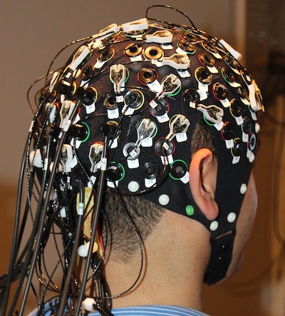
Simultaneous fNIRS-EEG acquisition for a visual task.
GFLI funding has allowed the CENIR to be equipped with an fNIRS acquisition device that comprises 16 sources and 16 detectors (NIRScoutTM, NIRx Medical Technologies, LLC) organized as source-detector optode pairs attached to optical fibers. The optodes are placed on the subject’s scalp and can be used simultaneously with EEG electrodes. The sources (electroluminescent diodes) emit near-infrared light with two different wavelengths (760 nm and 850 nm). The detectors receive the light that has travelled through the brain without being absorbed by biological tissues. The magnitude of the recorded signal then depends on the concentration in deoxy- and oxy-hemoglobin along the path length that the light has travelled in the cortex.
Functional NIRS enables to acquire high frequency (100 Hz) signals with 5 mm to 1-2 cm spatial resolution. However, due to strong light absorption in biological tissues, only the outermost cortex can be imaged and deep brain structures remain inaccessible.
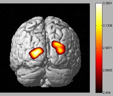
Activated brain areas during a visual task (example of deoxy-hemoglobin concentration map) obtained from fNIRS data processing.
The fNIRS technique is complementary to EEG and MEG. Activated brain areas, mapped on the scalp, can be compared with those obtained from simultaneous EEG recording. This allows one to assess the relationship between electromagnetic activity and hemodynamic signal, and to characterize brain networks with connectivity measures similar to those used in fMRI and EEG/MEG.